Medical Imaging
This is a live streamed presentation. You will automatically follow the presenter and see the slide they're currently on.
This is a live streamed presentation. You will automatically follow the presenter and see the slide they're currently on.
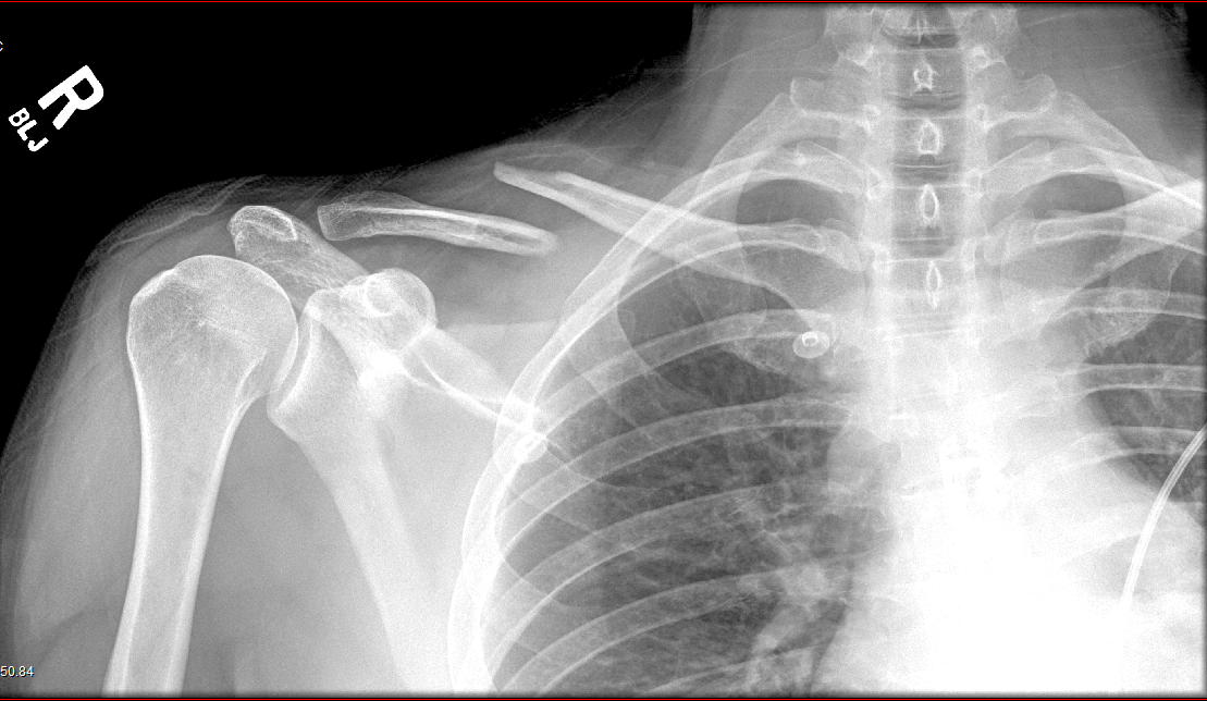
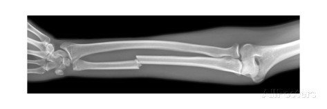
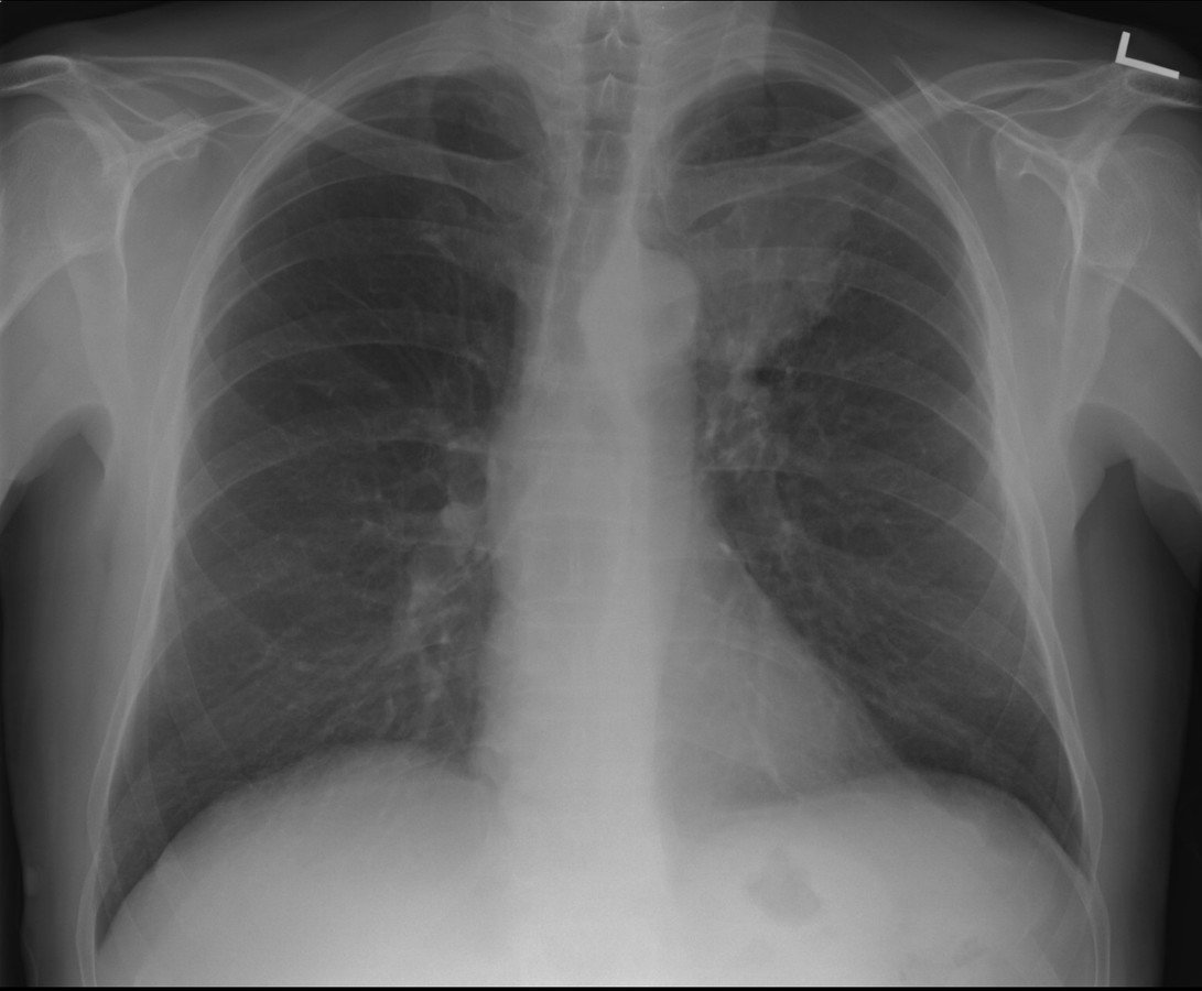
(X-Rays)
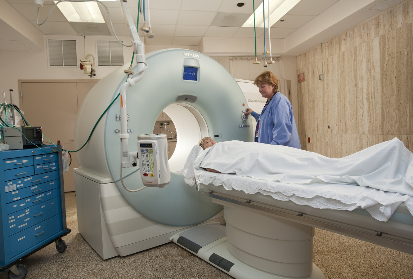
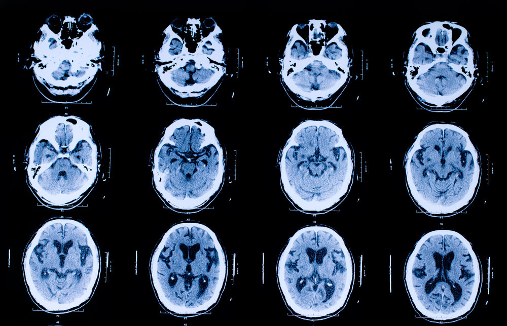
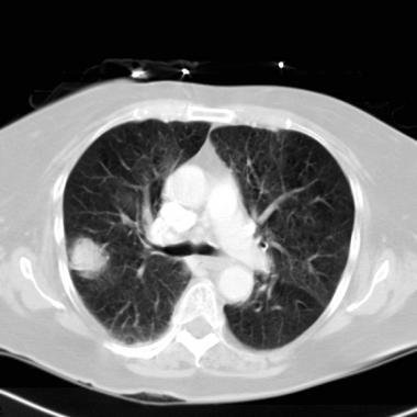
(CT Scan)
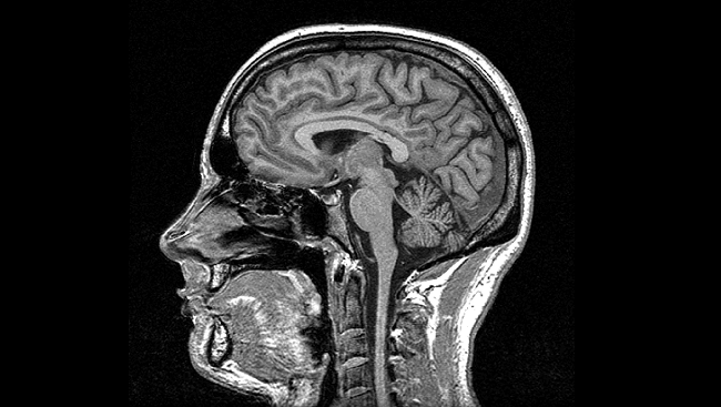
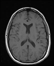
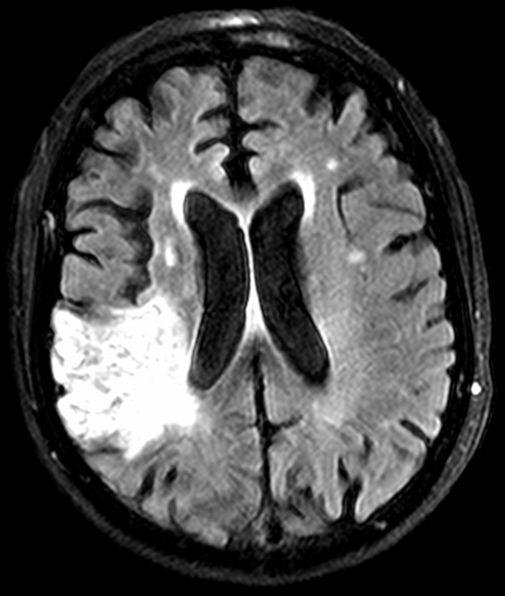
(MRI)