Cardiac Output by Echocardiography
Atul Jaidka
How to
SV = CSA (cm^2) x VTI (cm) = 0.785 x (LVOTd)^2 x (LVOT VTI)
CO = SV x HR
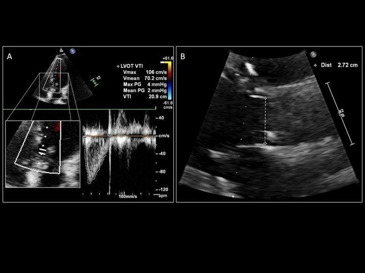
Sattin, Burhani, Jaidka, Arntfield. How I Do It: Echocardiographic-based Stroke Volume and Cardiac Output Determination. CHEST.
Pitfalls
LVOT VTI generated when the Doppler line of interrogation is (A) parallel to blood flow versus (B) off-axis by greater than 20˚
Sattin, Burhani, Jaidka, Arntfield. How I Do It: Echocardiographic-based Stroke Volume and Cardiac Output Determination. CHEST.
Pitfalls
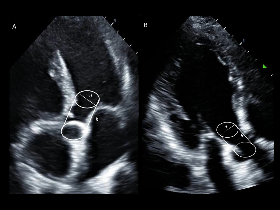
Difference between an LVOT VTI obtained from an (A) appropriate sample location in LVOT with aortic valve closure captured at the end of systole, and (B) a location too proximal in the LV cavity
Sattin, Burhani, Jaidka, Arntfield. How I Do It: Echocardiographic-based Stroke Volume and Cardiac Output Determination. CHEST.
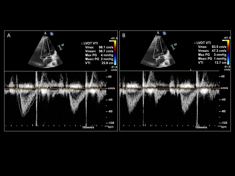
Pitfalls
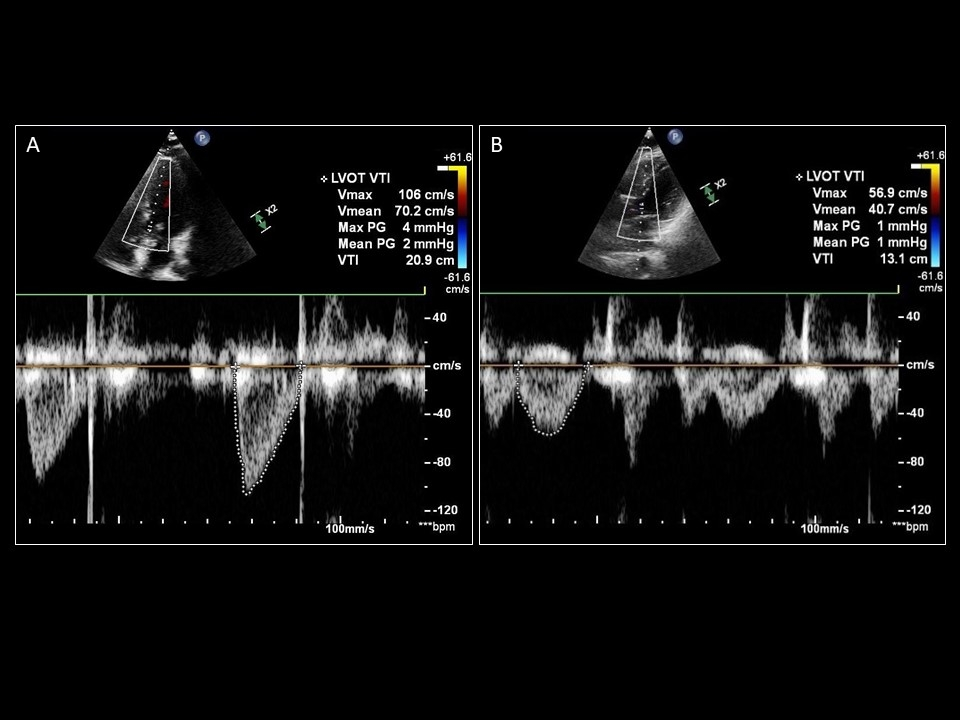
Demonstration of the significant difference in values obtained from over-tracing (A; 23.9cm) and under-tracing (B; 13.7cm) the same LVOT VTI.
Sattin, Burhani, Jaidka, Arntfield. How I Do It: Echocardiographic-based Stroke Volume and Cardiac Output Determination. CHEST.
Evidence
1. Stroke volume by echo predicts outcome
Heart and Soul Study
"Reduced stroke distance predicts HF hospitalization and mortality independent of clinical and other echocardiographic parameters among ambulatory adults with coronary artery disease."
https://www.sciencedirect.com/science/article/abs/pii/S089473171001093X
Evidence
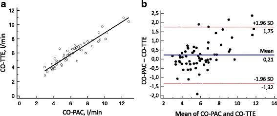
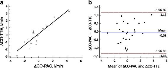
Many small studies show apparent correlation between Cardiac output by echo and Pulmonary Artery Catheter
https://www.ncbi.nlm.nih.gov/pmc/articles/PMC5465531/
Evidence
Systematic review of cardiac output measurements by echocardiography vs. thermodilution: the techniques are not interchangeable
(Intensive Care Med. 2016 Aug;42(8):1223-33. doi: 10.1007/s00134-016-4258-y. Epub 2016 Mar 1.)
- "24 studies comparing echocardiography with thermodilution as the clinical standard technique in measuring CO, but only two studies had a design that allowed a fully unbiased comparison of the two techniques"
- One these two studies assess precision of individual measures by both PAC and Echo
- Patient physiology can impact both measures (ie. TR and CO-TD)
- CO by doppler assumes LVOT is a cylinder but real world it is not
- "Current evidence does not support interchangeability between these techniques in measuring cardiac output. Thermodilution and echocardiography may be interchangeable in tracking directional changes in cardiac output"
https://pubmed.ncbi.nlm.nih.gov/26932349/
Take Home Points
- Cardiac output by echo is possible
- Important to know limitations in assessment
- Not enough data to support usage of CO in magnitude but possibly in trajectory