Quality Assurance Rounds
Echocardiolography | Atul Jaidka
Imaging
- TTE (pre-diagnosis/intervention)
- TEE (pre-diagnosis/intervention)
- Presenting both together, per valve
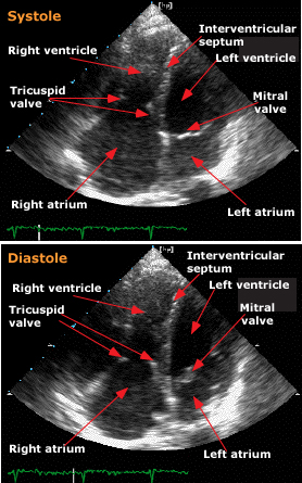
Ventricles
Left Ventricle
- Normal function, EF 63%
- Mildly dilated by volume
- D shaped appearance
TTE
Left Ventricle
- Normal function, EF 63%
- Mildly dilated by volume
- D shaped appearance
TTE
Right Ventricle
- Normal size and function
TTE
Valves
Aortic Valve
TTE
Aortic Valve
TEE
Aortic Valve
TEE
Aortic Valve
- Tricuspid and thickened valve
- RCC thickened and immobile
- Moderate regurgitation, no stenosis
TEE
Mitral Valve
TTE
Mitral Valve
TTE
Mitral Valve
TEE
Mitral Valve
TEE
- Immobile PMVL
- Mild thickening of subchordal apparatus
- Severe regurgitation, no stenosis
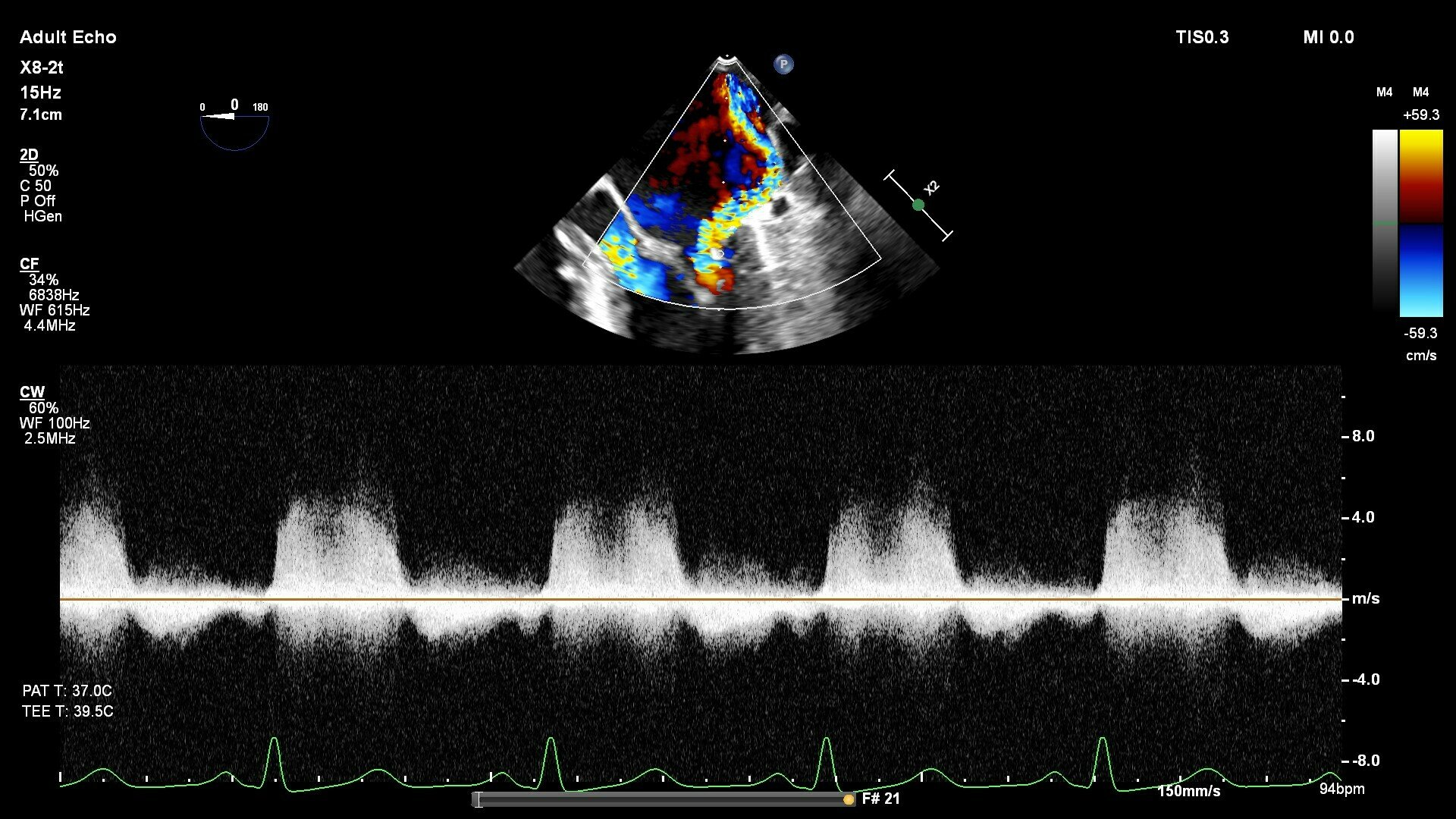
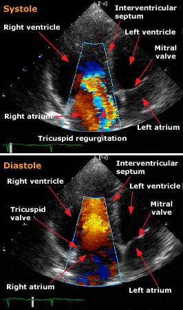
Tricuspid Valve
TTE
Tricuspid Valve
TEE
Tricuspid Valve
TEE
- Restricted motion of anterior leaflet
- No thickening of subchordal apparatus
- Severe regurgitation, no stenosis
Pulmonary Valve
TTE
- Mildly reduced excursion of leaflets
- Mild stenosis
- Moderate regurgitation
Summary
- Normal LV and RV function
- Immobile posterior mitral valve leaflet
- Severe MR
- Aortic valve with thickened cusps and restricted RCC
- Moderate AR
- Restricted motion of anterior tricuspid leaflet
- Severe TR
- Thickened pulmonary valve with reduced excursion
- Mild PS, Moderate PR
- No shunt seen by 2D, colour Doppler or bubble study
Differential
-
Findings are consistent with post-inflammatory changes
- Differential:
-
Rheumatic heart disease (most common)
-
SLE, other autoimmune conditions
-
Radiation exposure/injury
-
Carcinoid heart disease
-
Drug-induced valvulopathy (eg. ergot alkaloids)
-
Endocarditis (infective/non-infective
-
Carcinoid Heart Disease
UTD
Carcinoid Heart Disease
UTD
Carcinoid Heart Disease
Rheumatic Heart Disease
Rheumatic Heart Disease
UTD
Rheumatic Heart Disease
UTD