SVT Case
Atul Jaidka
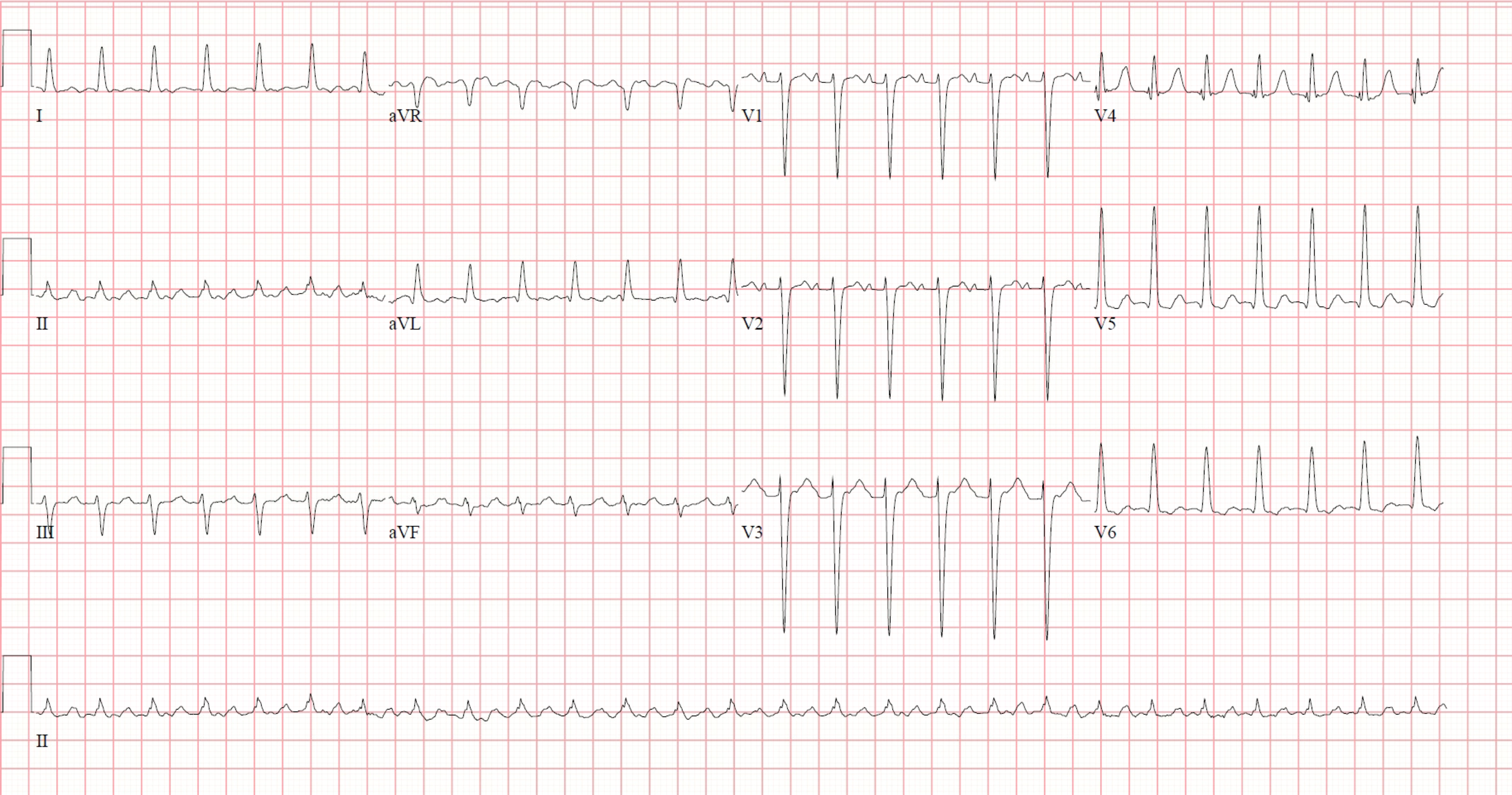
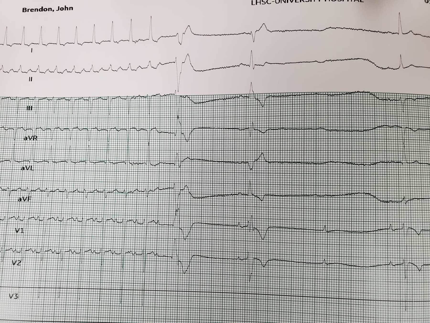
SVT
Adenodine
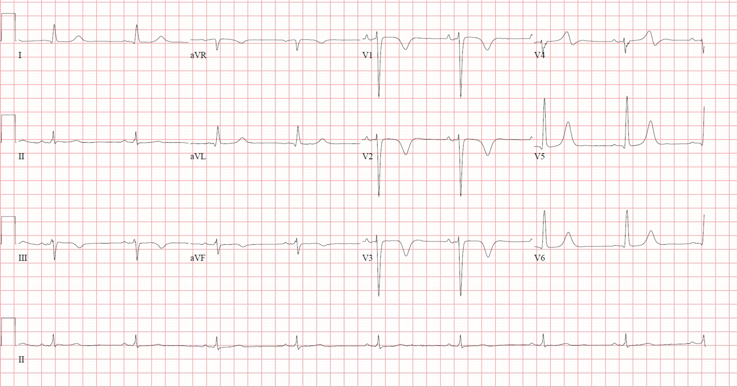
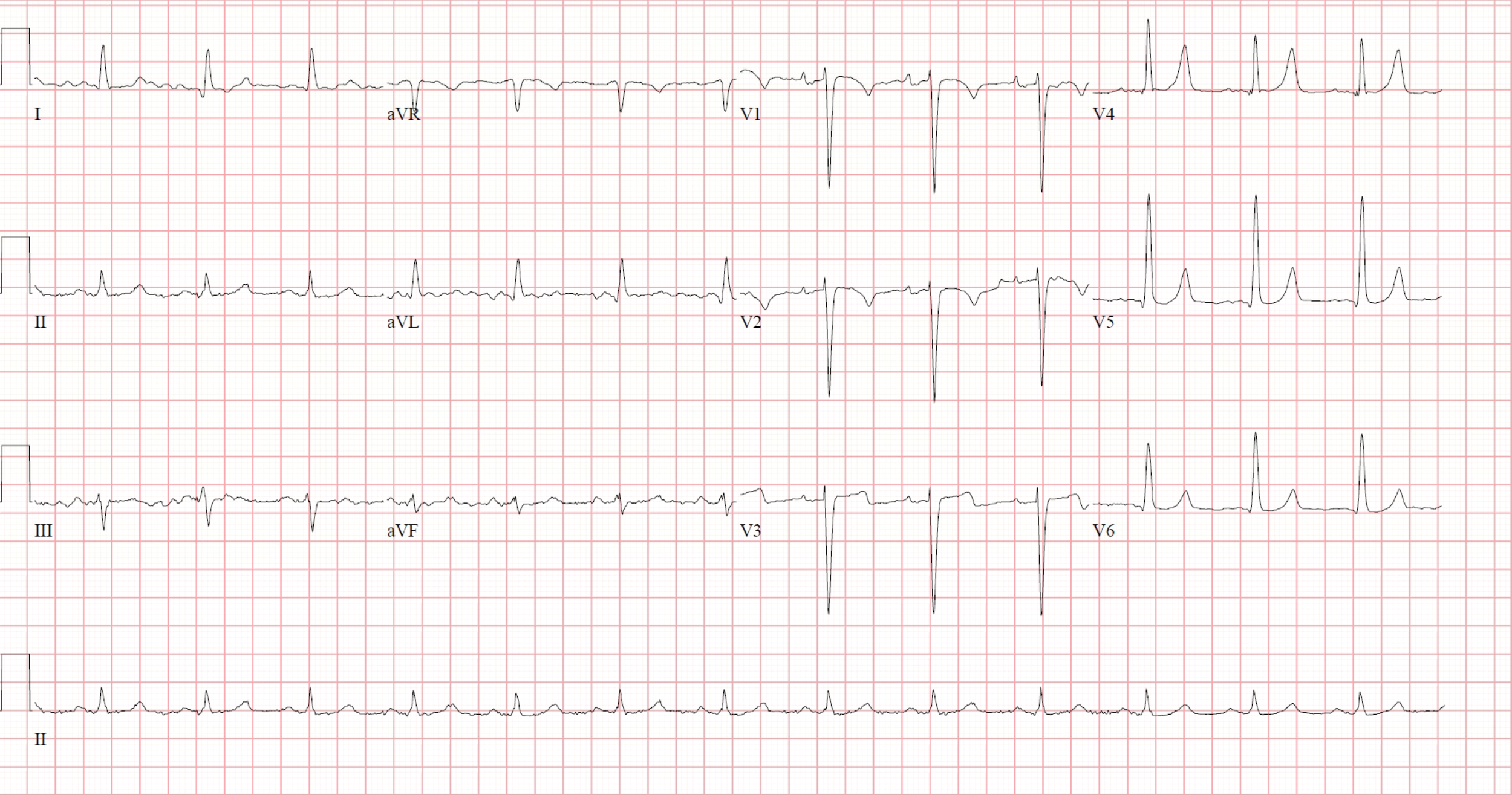
Sinus rhythm
Echo
Left ventricle is mildly dilated.
Left ventricular systolic function is severely reduced (Ejection Fraction = 25-30%).
Severe global hypokinesis of the left ventricle.
No LV thrombus.
Left atrium is moderately dilated.
Mild mitral regurgitation.
Clinical
- Refractory SVT despite metoprolol 100mg BID and repeated verapamil and adenosine doses
Ablation
An electrophysiology study was performed which demonstrated the presence of eccentric atrial activation with ventricular pacing. Using extrastimuli, AVRT was induced. Hence, using ICE and fluoroscopic guidance, a transeptal puncture was performed with a BRK-1 needle though an SR-0 sheath. This was quite challenging as the heart was rotated (Needle positioned at 7'o'clock then counter-clocked till seen on Fossa via ICE) but was performed successfully. Heparin was given once the wire was confirmed to be within the LA.
The left lateral pathway was targeted during ventricular pacing. Radiofrequency ablation was delivered using an ST-SF D curve, irrigated catheter. The left lateral pathway was successfully ablated at 3 o’clock on the MV annulus. Testing 20 minutes after ablation revealed no dual node physiology, echo beats, or inducible SVT. The sheaths and catheters were removed and the ICE demonstrated a stable pericardial effusion, likely from his HF.
Sinus rhythm post ABlation