AVANITHK.COM


Only public university in the Boston area

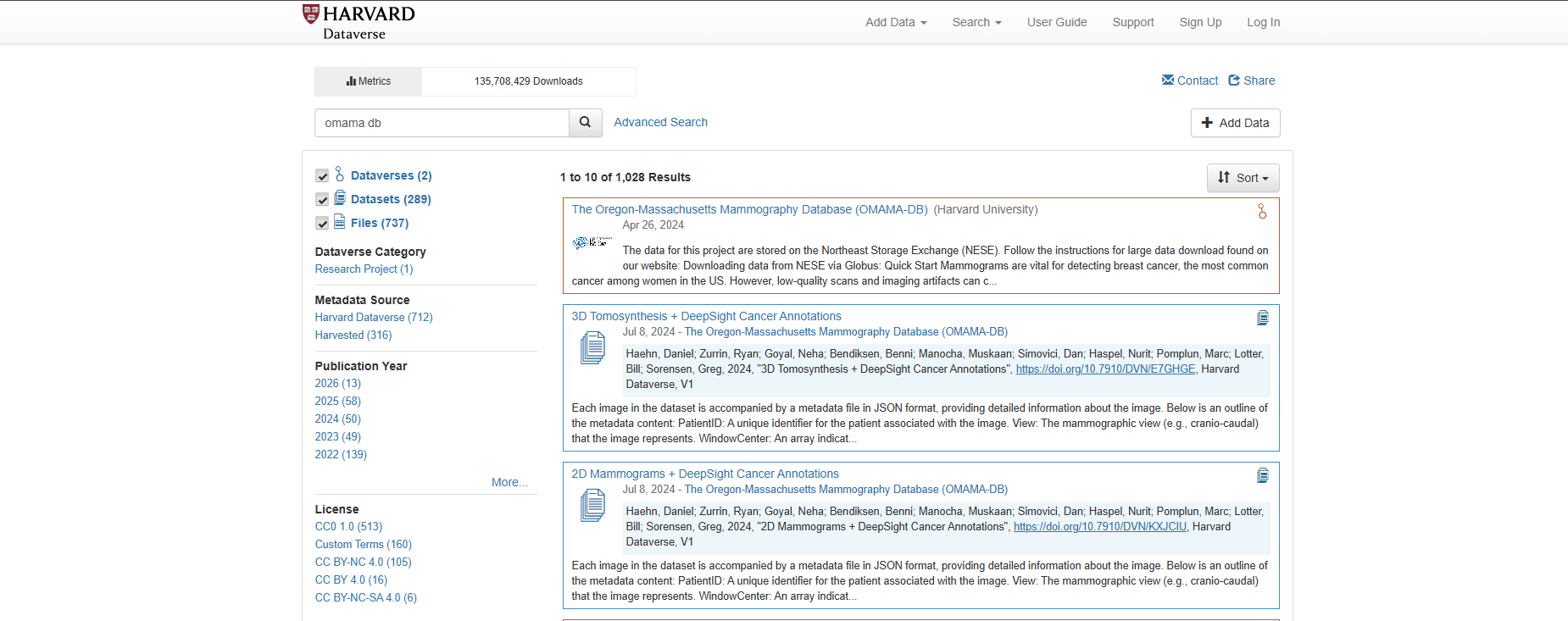
THE OREGON-MASSACHUSETTS MAMMOGRAPHY DATABASE
November 9, 2026
OMAMA-DB

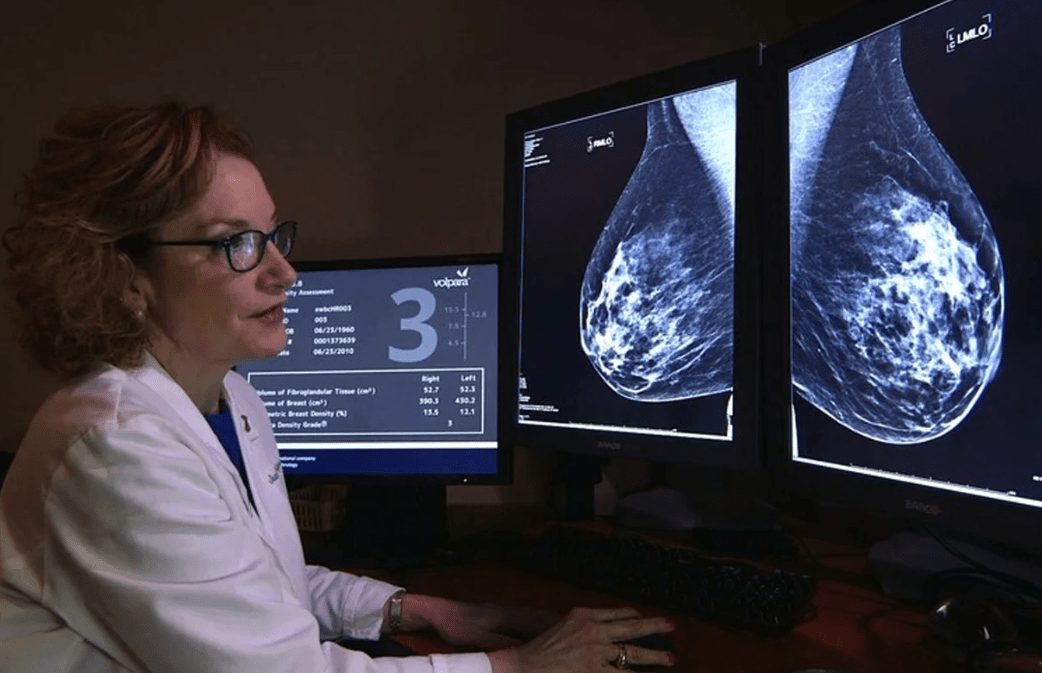
Breast cancer is the most common cancer among women worldwide.


2D
256x256.tar(3.0 GB)
1024x1024.tar(97.8 GB)
Original_2d.tar(741.3 GB)
512x512.tar(23.4 GB)

3D
3d.tar(7.9 TB)
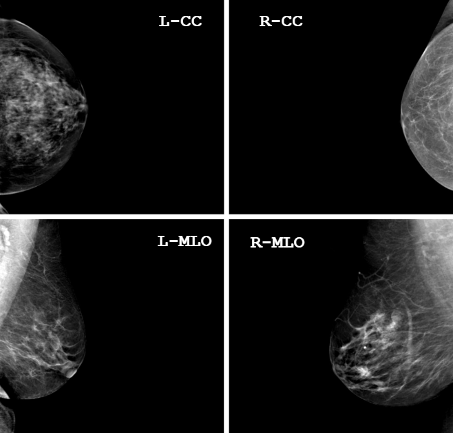
4-Views
{
Patient ID: DHVGGTHM5AB2
View: cranio-caudal
WindowCenter: [2575.0, 2653.0, 2479.0]
WindowWidth: [900.0, 750.0, 1050.0]
WindowCenterWidthExplanation: ["NORMAL", "HARDER", "SOFTER"]
ImagerPixelSpacing: [0.094090909, 0.094090909]
ImageLaterality: "R",
coords: [401, 112, 498, 219]
score: 0.8513884544372559,
label: IndexCancer
}
JSON File :
| Dataset | Public Images |
|---|---|
| DDSM | 10,480 |
| MIAS | 322 |
| INbreast | 410* |
| CMMD | 5,202 |
| VinDr-Mammo | 20,000 |
| OPTIMAM | 3,072,878* |
| EMBED | 480,323* |
| OMAMA-DB (Ours) | 231,080 |
Dataset Comparison
Download Stats
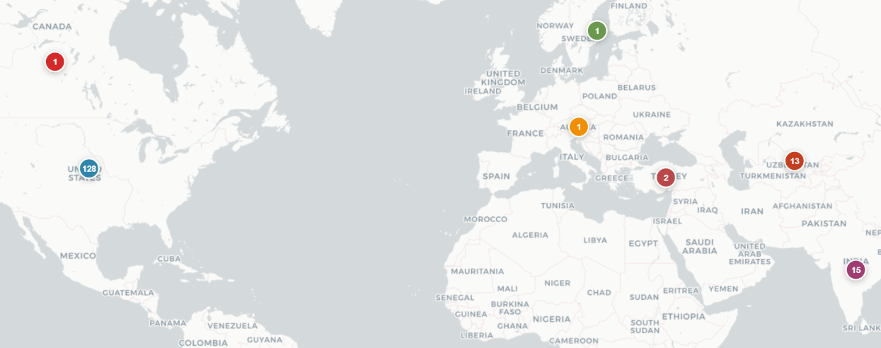
Global Usage
- 65% of downloads from USA
- 79% of downloads are 2D
- Used Worldwide
Top users:
- Students
- Researchers
- Universities
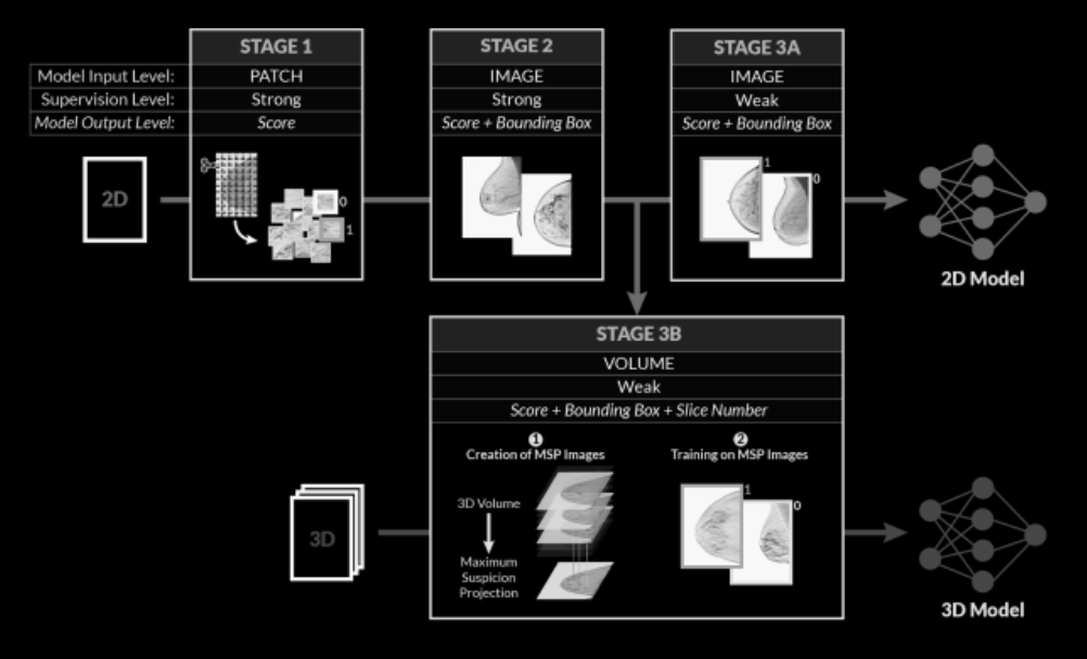
Dataset Creation
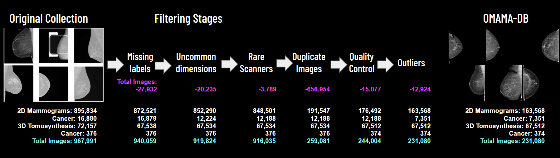
Dataset Creation
NonCancer
IndexCancer
PreIndexCancer
Laterality : L/R
Dataset Creation
46 x 56 px
512 x 564 px
Dataset Creation
Hologic, lnc
GE Medical Systems
Dataset Creation
Multiple Views
Duplicate Studies
Dataset Creation
Corrupted Dicom Headers
Missing Pixel Data
Dataset Creation


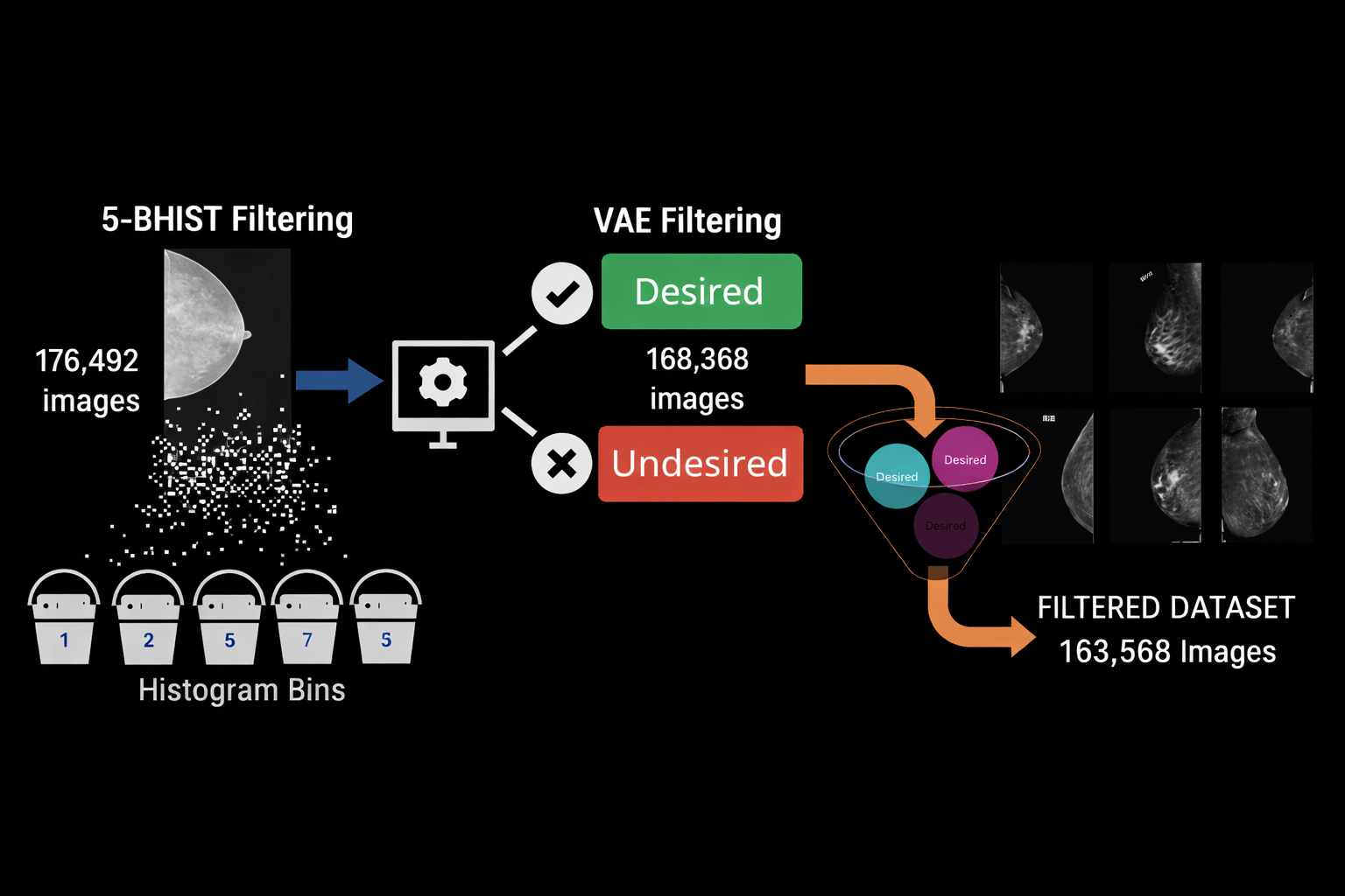
5-BHIST Filtering
VAE Filtering
Outlier Detection (ODM)
Medical Imaging with Deep Learning 2023
Published in :
Dataset Creation
Dataset Creation
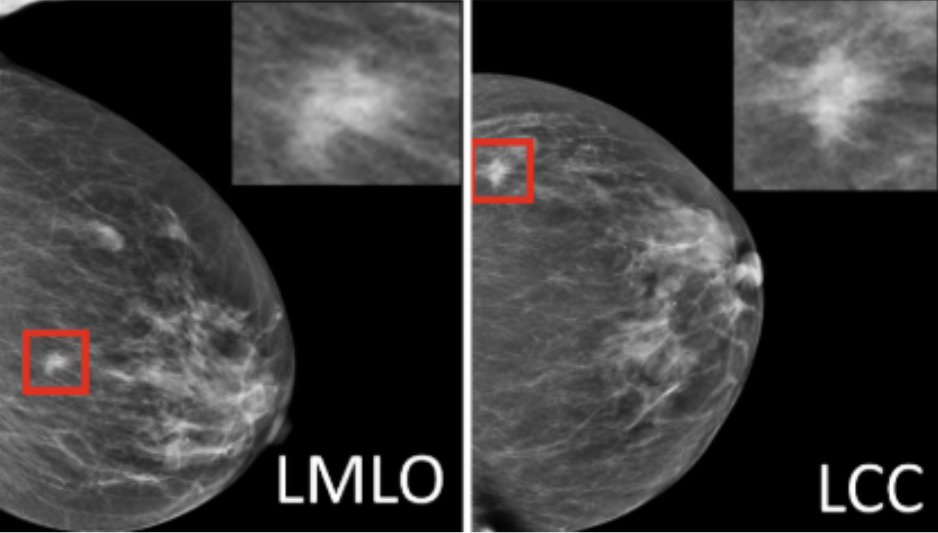
{
Patient ID: DHVGGTHM5AB2
View: cranio-caudal
WindowCenter: [2575.0, 2653.0, 2479.0]
WindowWidth: [900.0, 750.0, 1050.0]
WindowCenterWidthExplanation: ["NORMAL", "HARDER", "SOFTER"]
ImagerPixelSpacing: [0.094090909, 0.094090909]
ImageLaterality: "R",
coords: [401, 112, 498, 219]
score: 0.8513884544372559,
label: IndexCancer
}
JSON File :
Deepsight

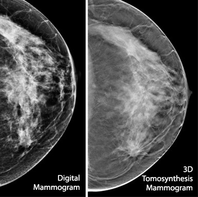
Visual exploration of mammography dataset
Tasks
T1: Identify artifacts, low-quality images, and outliers.
Tasks
T1: Identify artifacts, low-quality images, and outliers.
T2: Compare automated annotations with pathology-based labels.


pathology-based labels
NonCancer, PreIndexCancer, IndexCancer
VS
Tasks
T1: Identify artifacts, low-quality images, and outliers.
T2: Compare automated annotations with pathology-based labels.
T3: Explore structure across image types and acquisition context.
Tasks
T1: Identify artifacts, low-quality images, and outliers.
T2: Compare automated annotations with pathology-based labels.
T3: Explore structure across image types and acquisition context.
T4: Design a generic visual analytics approach for image + metadata + AI labels.


Tasks
T1: Identify artifacts, low-quality images, and outliers.
T2: Compare automated annotations with pathology-based labels.
T3: Explore structure across image types and acquisition context.
T4: Design a generic visual analytics approach for image + metadata + AI labels.


100 + FIELDS


2D Multiview
256x256.tar(655.0 MB)
1024x1024.tar(9.7 GB)
Original_2d.tar(74.0 GB)
512x512.tar(2.4 GB)

100 + FIELDS


mpsych.org/omama