BIOSC 1540: L09A (Structural biology)
This is a live streamed presentation. You will automatically follow the presenter and see the slide they're currently on.
This is a live streamed presentation. You will automatically follow the presenter and see the slide they're currently on.
Computational Biology
(BIOSC 1540)
Mar 11, 2025
Lecture 09A
Structural Biology
Foundations
Assignments
Quizzes
CBits
Supplementary material is available to read; not required, but recommended
At the foundation of biological processes lie atoms and their interactions
Structural biology studies the 3D shapes of biological macromolecules and how these shapes relate to function
Why study structure?
Primary Goal: To understand how molecular machines in cells work by deciphering their atomic arrangements.
CRISPR-Cas9
COVID-19 treatments
High-throughput sequencing
Innovation and biotechnology depend on molecular understanding
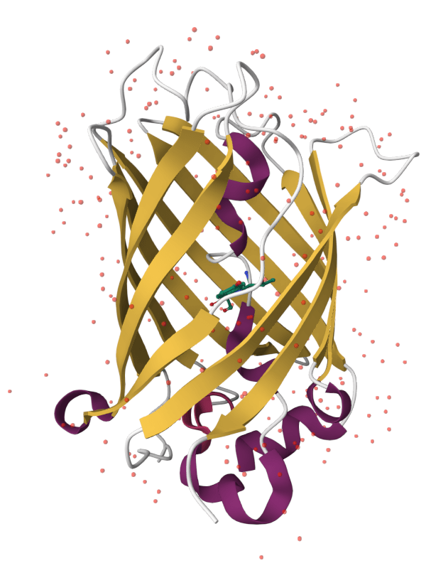
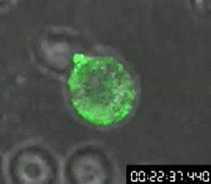
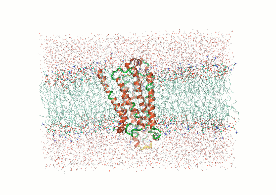
Alex's research example: Engineering green fluorescent protein with Dr. Rosenbaum and Dr. Carlson
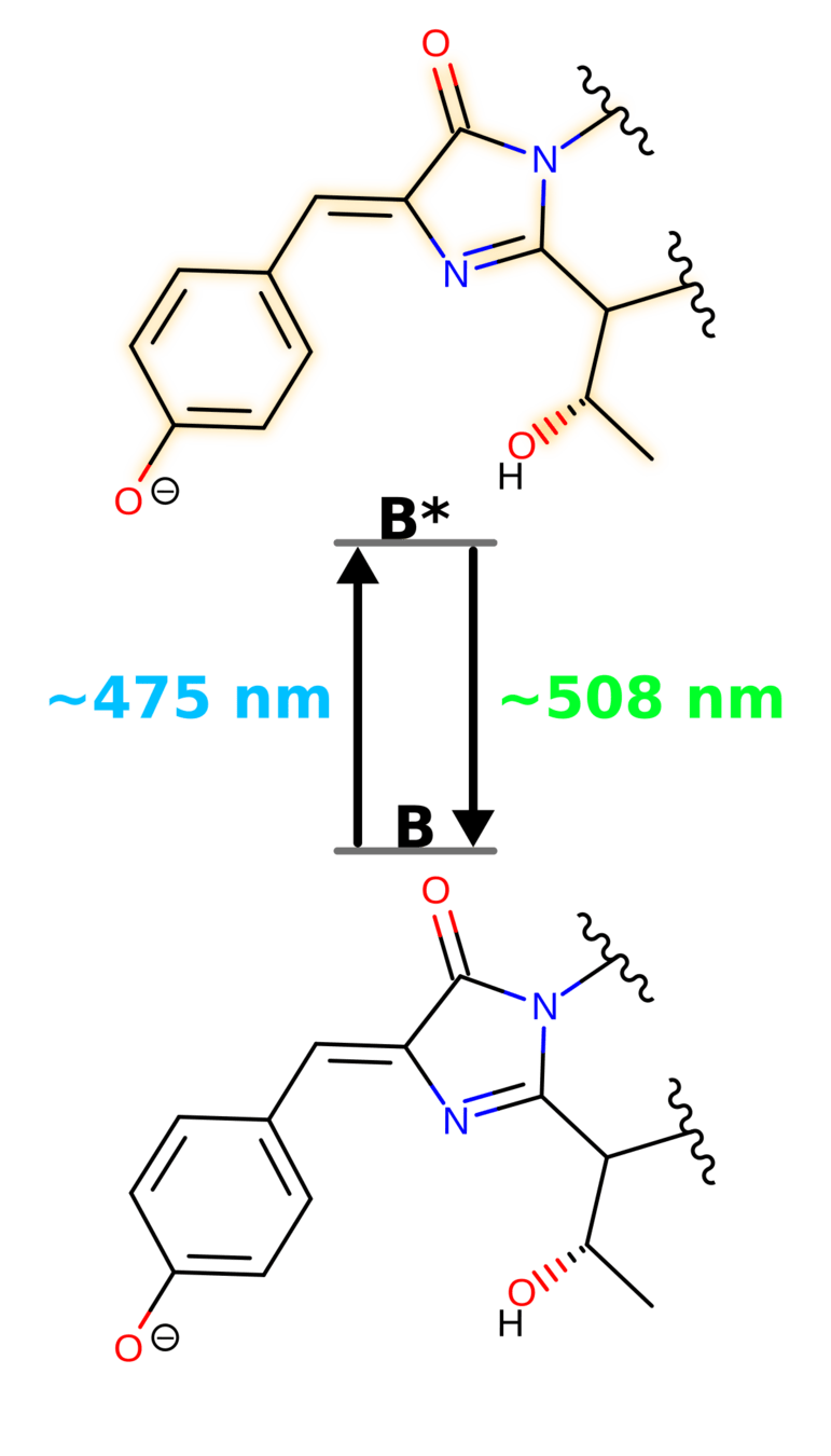
Enhanced GFP (eGFP) absorbs violet/blue light (400 - 490 nm) and emits green light ~507 nm
Track molecules by adding it as a tag
First video of cellular transfer of HIV
Differentiate cells with GFP variants
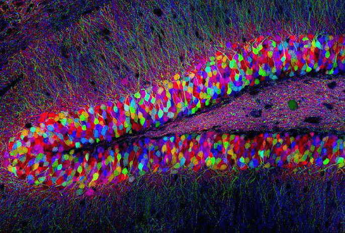
Multicolored GFPs used to map mouse brain
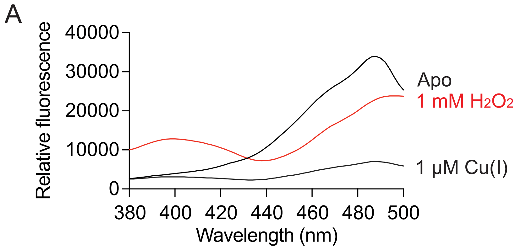
Redox potentials indicate a solution's tendancy to gain or lose electrons
For example, mitochondria are highly reducing with a redox potential around -0.36 V
Reduction: NAD+ to NADH
Oxidation: NADH to NAD+
Hanson, G. T., et al. (2004). Journal of Biological Chemistry, 279(13), 13044-13053. DOI: 10.1074/jbc.M312846200
-0.310 V
-0.275 V
-0.240 V
Fluorescence ratio after 400/488 nm excitation correlated to redox potential of roGFP2 environment
Hanson, G. T., et al. (2004). Journal of Biological Chemistry, 279(13), 13044-13053. DOI: 10.1074/jbc.M312846200
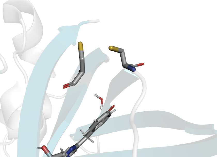
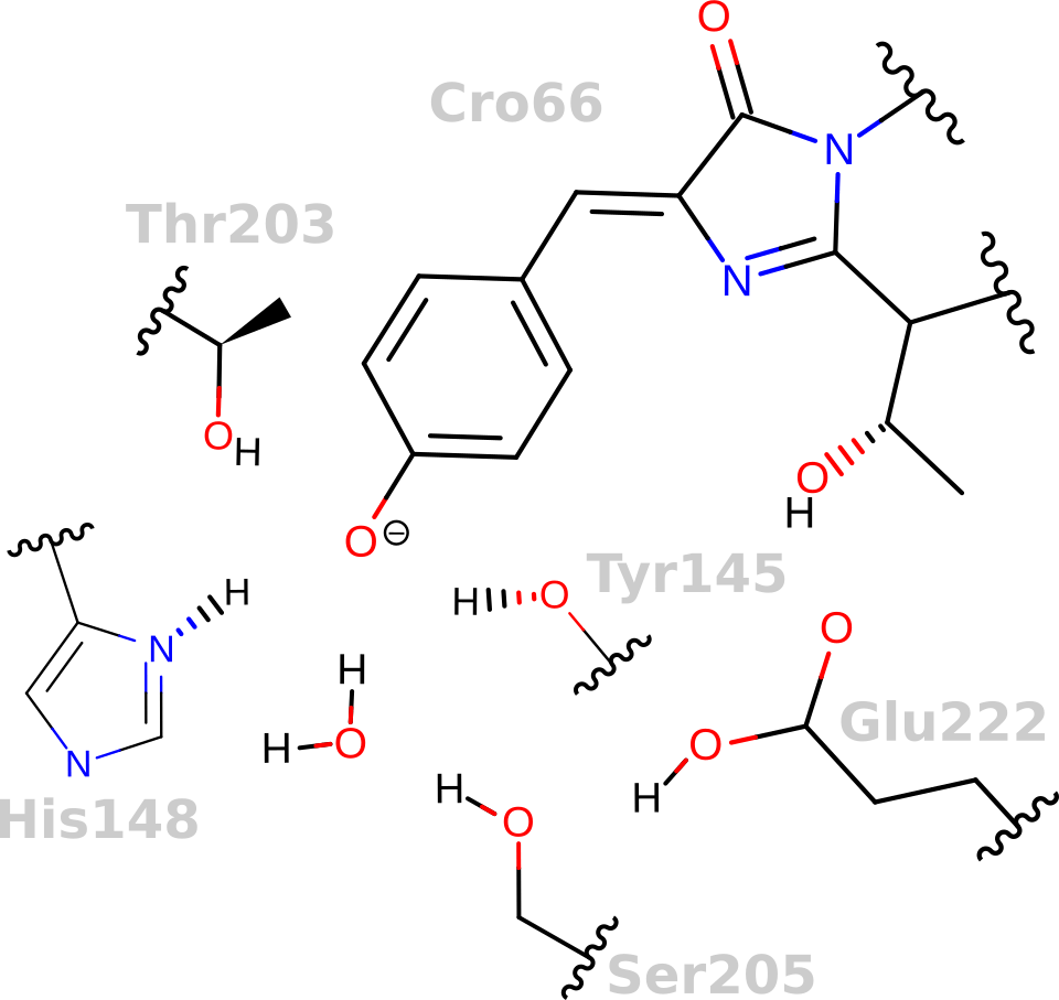
PDB ID: 2Y0G
PDB ID: 1JC0
147
204
CRO
147
204
CRO
(Contains S65T "enhanced" mutation)
Wild type
roGFP2
Hanson, G. T., et al. (2004). Journal of Biological Chemistry, 279(13), 13044-13053. DOI: 10.1074/jbc.M312846200
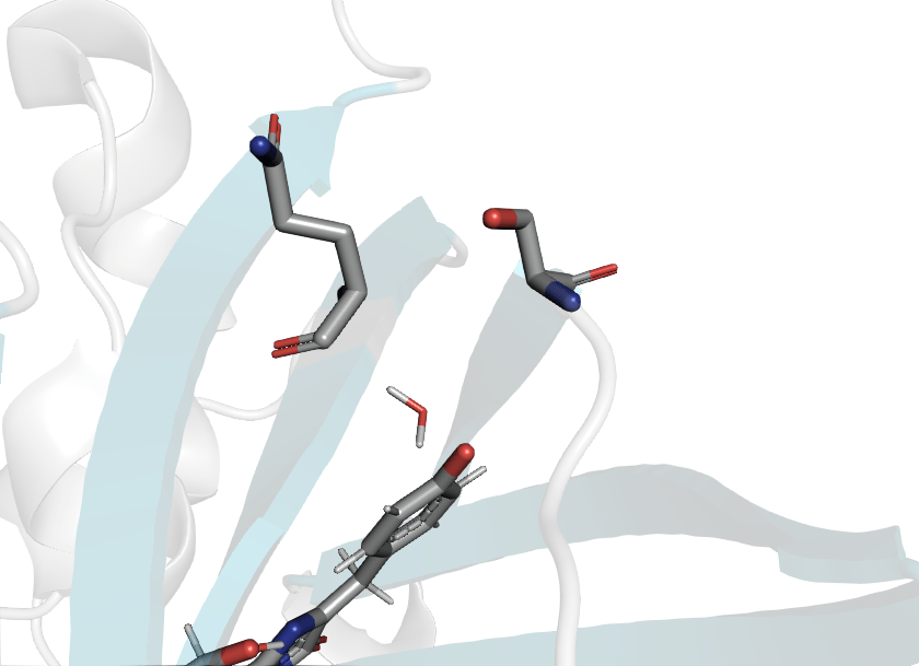
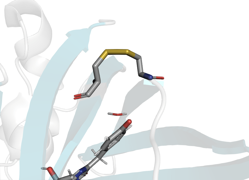
Reduced
Oxidized
PDB ID: 1JC0
PDB ID: 1JC1
(Contains S65T "enhanced" mutation.)
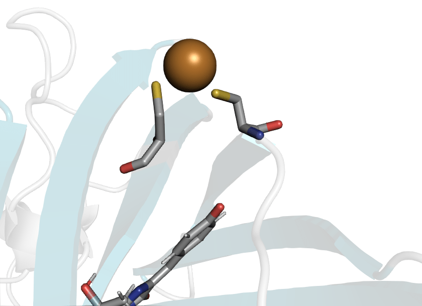
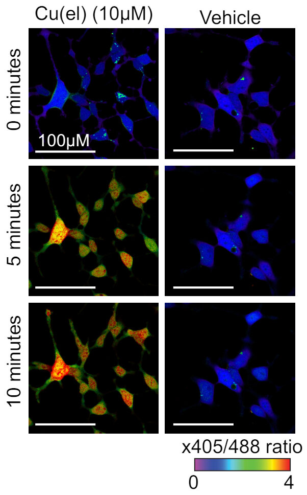
roGFP2 can bind Cu(I) to CYS147 and CYS204
Computational question: How does Cu(I) binding quench roGPF2 florescence?
roGFP2 will also change fluorescence in a different way when copper is present
When the chromophore has increased flexability, it will de-excite through vibrations instead of emitting photons
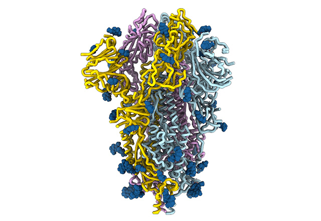
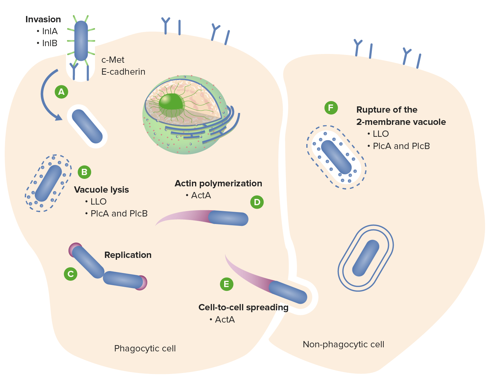
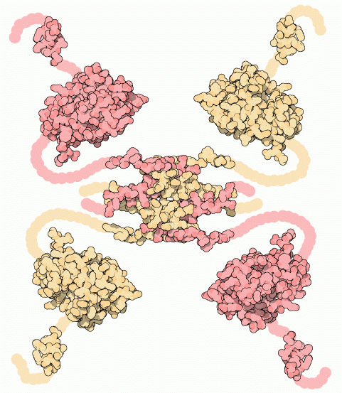
Alex's research example: Listeria monocytogenes with Dr. Cahoon
Lm is a gram-positive bacteria responsible for listeriosis, a foodborne illness
Agbavor, C.; et al. DOI: 10.1128/mbio.00743-24
A key step in the Lm life cycle is escaping vacuoles and continue infecting
PrsA2
PrsA2
LLO
Lm secretes a pore-forming, cholesterol-dependent toxin called listeriolysin O (LLO) to escape vacuoles and infect cells
Agbavor, C.; et al. DOI: 10.1128/mbio.00743-24
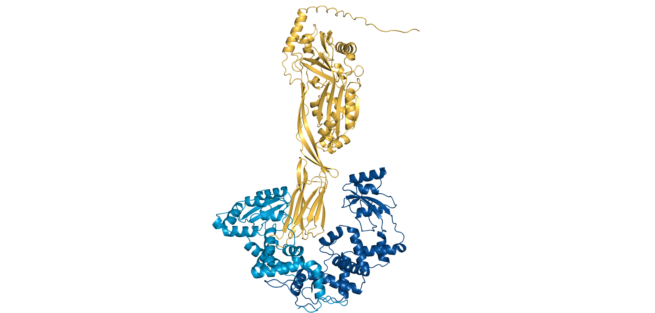
The Cahoon lab (alongside several collaborators) demonstrated that PrsA2 (a chaperone) regulates LLO activity through a pH-dependent mechanism
At pH 7, PrsA2 remains bound to LLO, preventing it from forming pores. At pH 5, PrsA2 releases LLO to escape acidic vacuoles
This is a new project, so we do not know yet!
Are PrsA2-LLO interactions destabilized in acidic (i.e., pH 5) environments? If so, how?
PrsA2
PrsA2
LLO
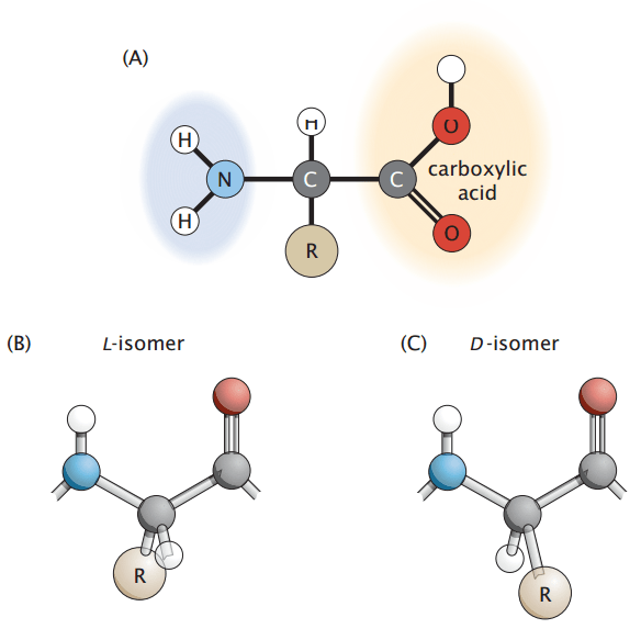
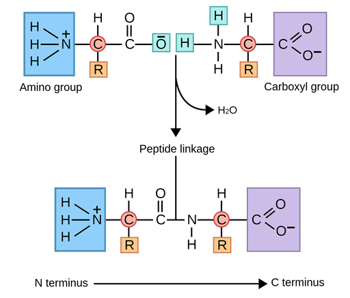
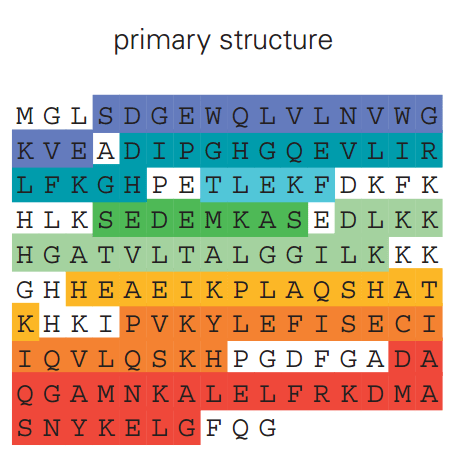
All proteins are composed of smaller molecules called amino acids, which are linked together in specific sequences.
Each amino acid contains a central carbon (alpha carbon) bonded to an amino group (NH₂), a carboxyl group (COOH), a hydrogen atom, and a variable side chain known as the R-group.
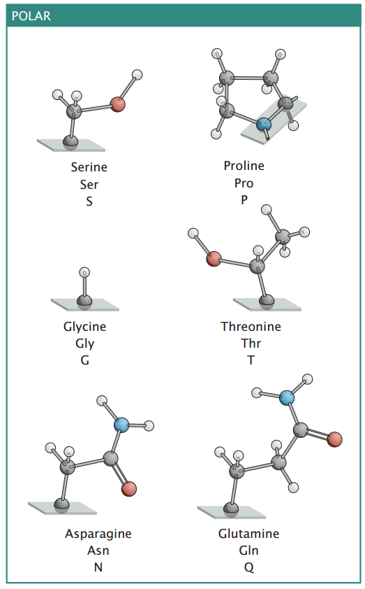
Polar amino acids have side chains that can form hydrogen bonds, making them hydrophilic
You will not be tested on your amino acid abbreviations
Polar amino acids contribute to protein solubility and help stabilize secondary and tertiary structures through hydrogen bonding.
Many polar amino acids are involved in enzymatic activity, facilitating catalytic reactions by stabilizing transition states or interacting with substrates.
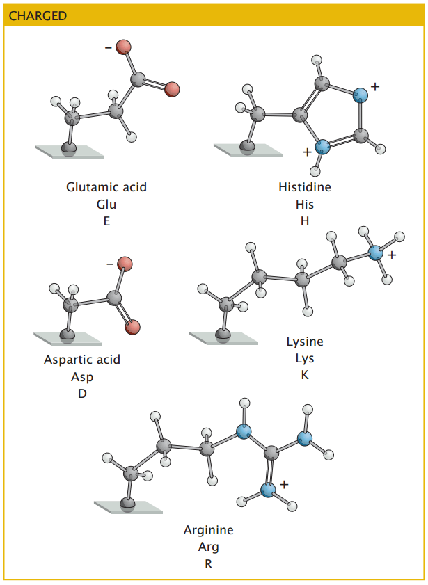
Acidic amino acids carry negative charges and participate in ionic interactions that stabilize protein structures
Charged amino acids contribute to protein folding by forming salt bridges, which enhance stability.
The cellular environment's pH can influence these amino acids' charge state, affecting protein conformation and function.
Basic amino acids carry positive charges and frequently interact with negatively charged molecules like DNA and phospholipids.
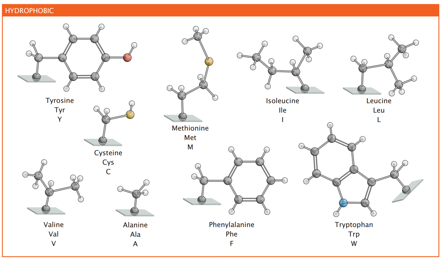
These amino acids are often found in the interior of globular proteins, stabilizing protein structure by minimizing exposure to water
Aromatic nonpolar amino acids participate in stacking interactions, influencing protein stability and ligand binding
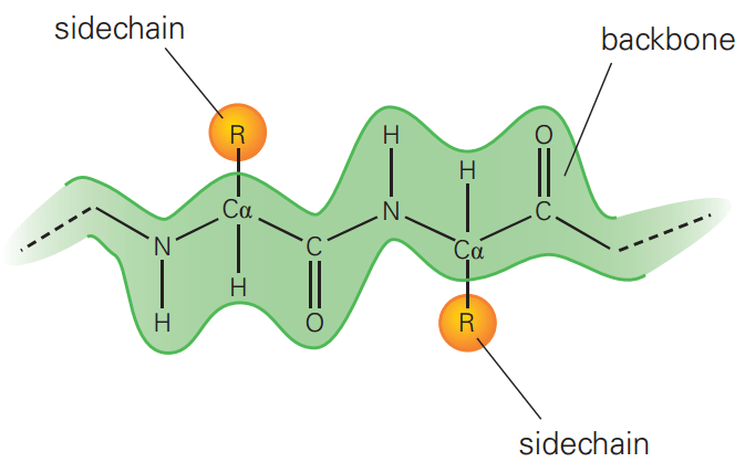
The primary structure of a protein is the linear sequence of amino acids, held together by covalent peptide bonds
The primary structure alone does not reveal the protein's functional form or activity
While the primary sequence is critical, the folding process may also depend on cellular factors (e.g., chaperones)
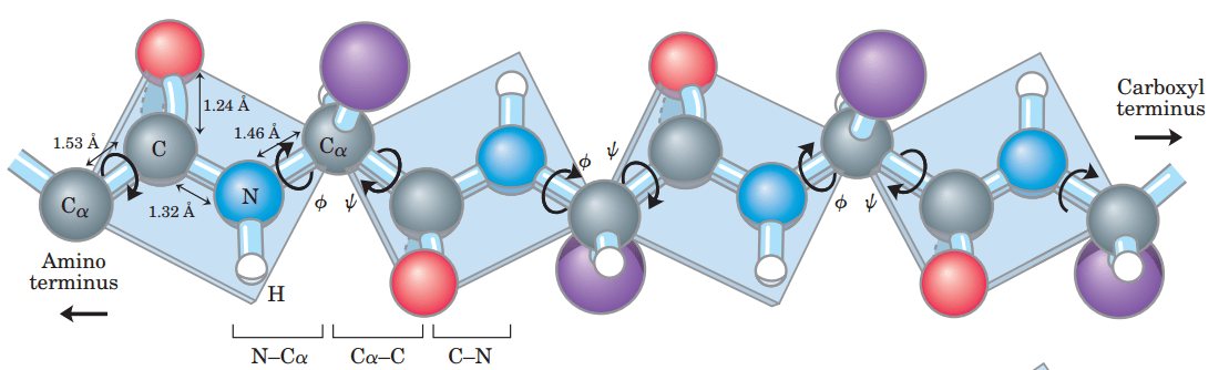
Proteins are flexible due to rotation around specific backbone bonds: the phi (Φ) and psi (Ψ) angles.
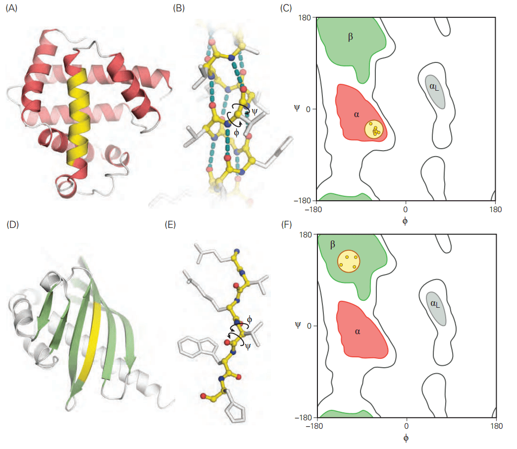
Not all angle combinations are allowed due to steric hindrance—this is visualized in a Ramachandran plot, which maps permitted conformations.
Secondary structures refer to regularly repeating local conformations of the polypeptide backbone.
These structures help proteins achieve compact and stable folding while maintaining flexibility for function.
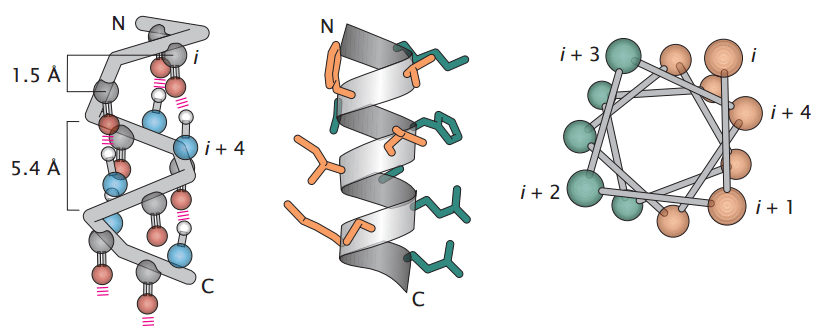
An alpha-helix is a right-handed coil with 3.6 amino acids per turn, stabilized by hydrogen bonds between the backbone carbonyl oxygen and the amide hydrogen of a residue four positions ahead.
Side chains project outward, allowing interactions with the surrounding environment.
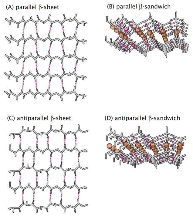
Beta-sheets consist of extended polypeptide strands aligned side by side, stabilized by hydrogen bonds between backbone atoms of adjacent strands.
Strands can be parallel (N-to-C direction aligned) or antiparallel (N-to-C in opposite directions), with antiparallel sheets being more stable.
Side chains alternate above and below the sheet, affecting interaction and stability.
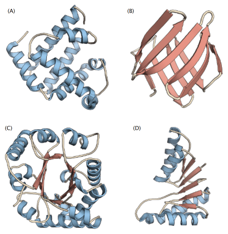
The tertiary structure refers to the complete 3D shape of a single polypeptide chain
Tertiary structures reveal active sites or binding pockets where catalysis or molecular interactions occur
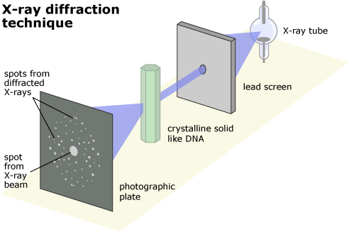
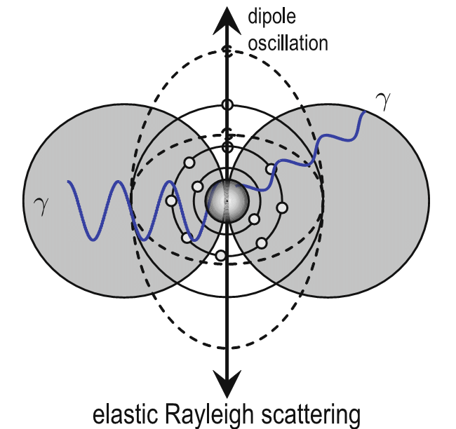
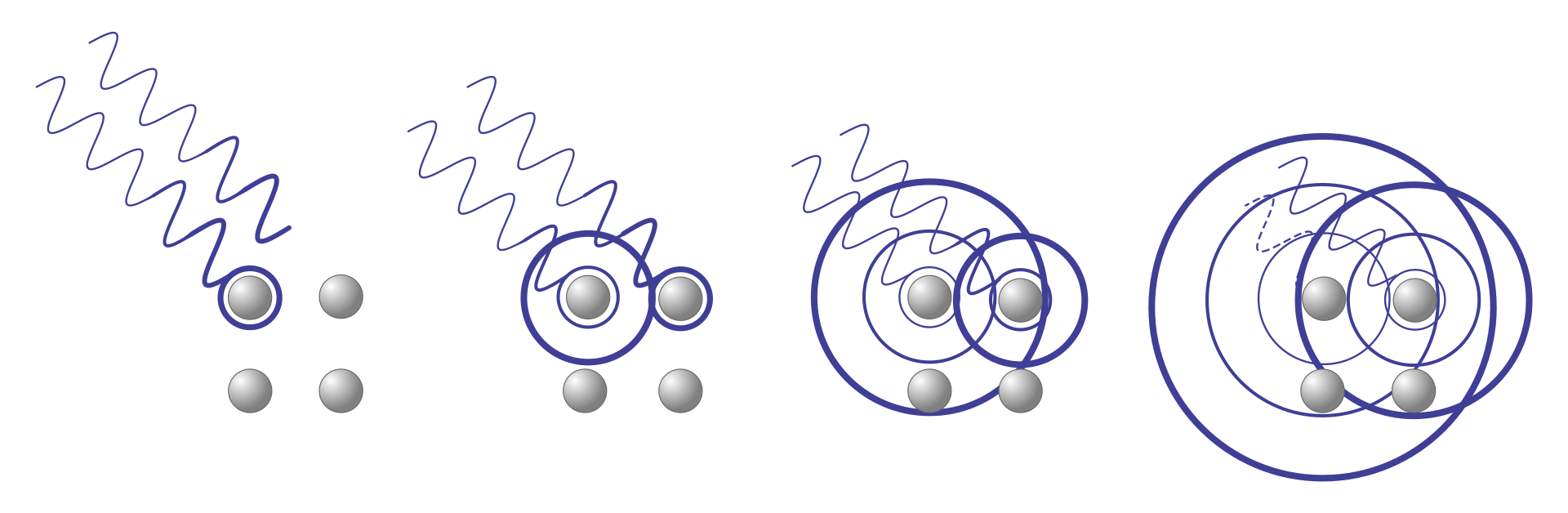
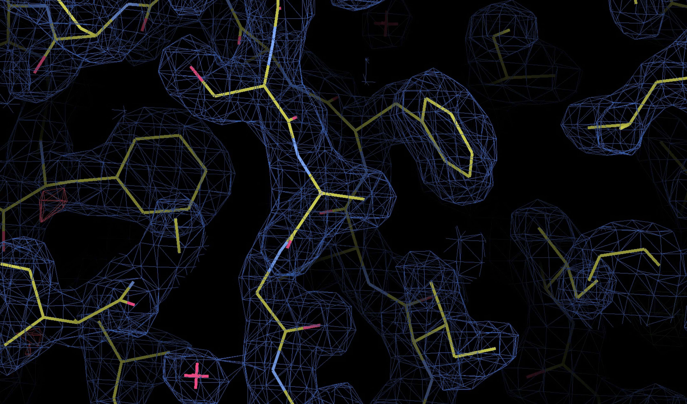
Basic Principle: Photons scatter when they interact with other particles
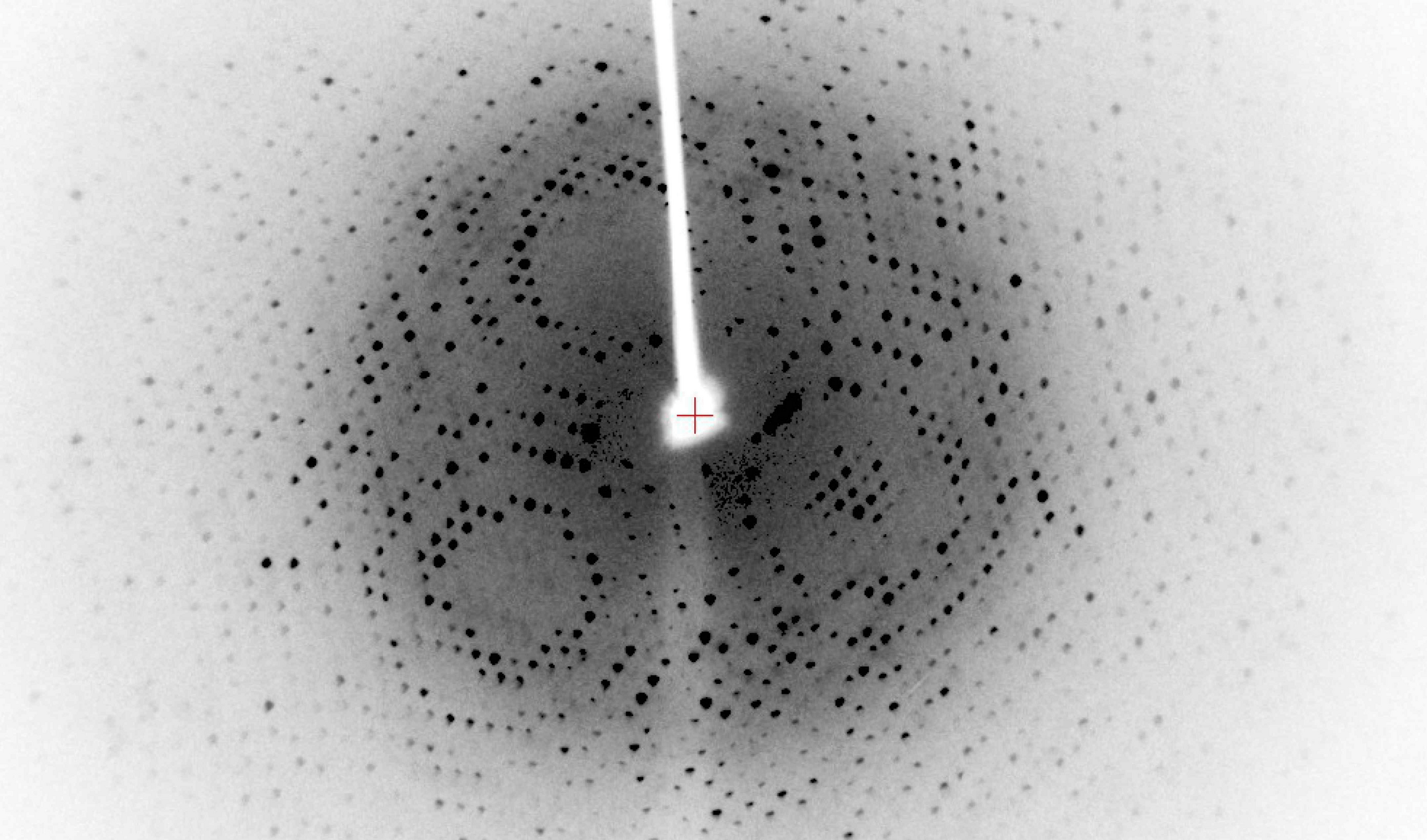
The scattered X-rays form a diffraction pattern unique to the crystal
Probe: Photon (carrier of electromagnetic radiation)
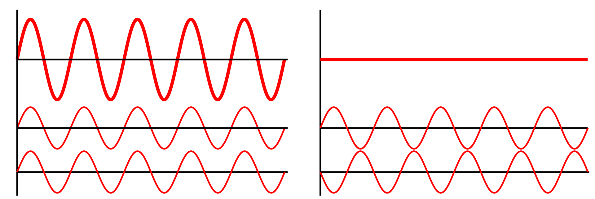
What happens when two waves overlap?
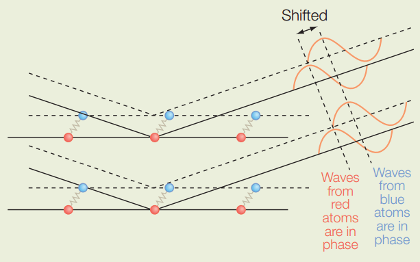
If wavelengths are similar and in phase, they constructively interfere
If waves are out of phase, they deconstructively interfere
If wavelengths are similar and in phase, they constructively interfere and form spots based on atom type and distance
The spots on the detector represent the reflections of the scattered X-rays
The diffraction pattern does not directly show the atomic positions, but provides the data needed to infer the electron density
The 3D electron density map reveals the distribution of electrons in the crystal, indicating where atoms are located
The electron density map is interpreted by fitting atomic models (e.g., amino acids for proteins) into the density
Low-resolution data make it difficult to assign atomic positions precisely, leading to uncertainty in the model
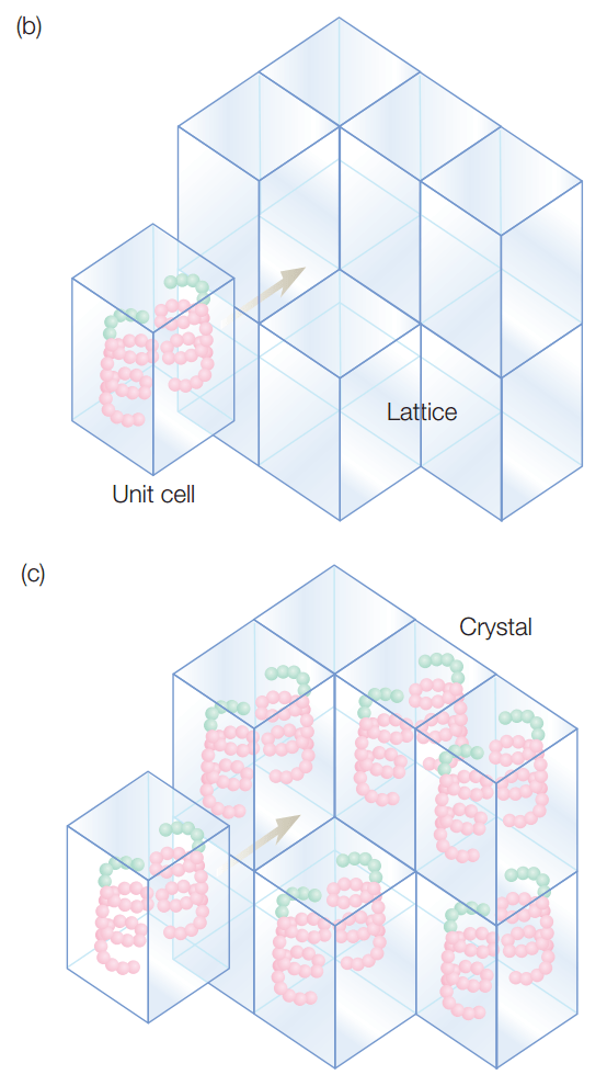
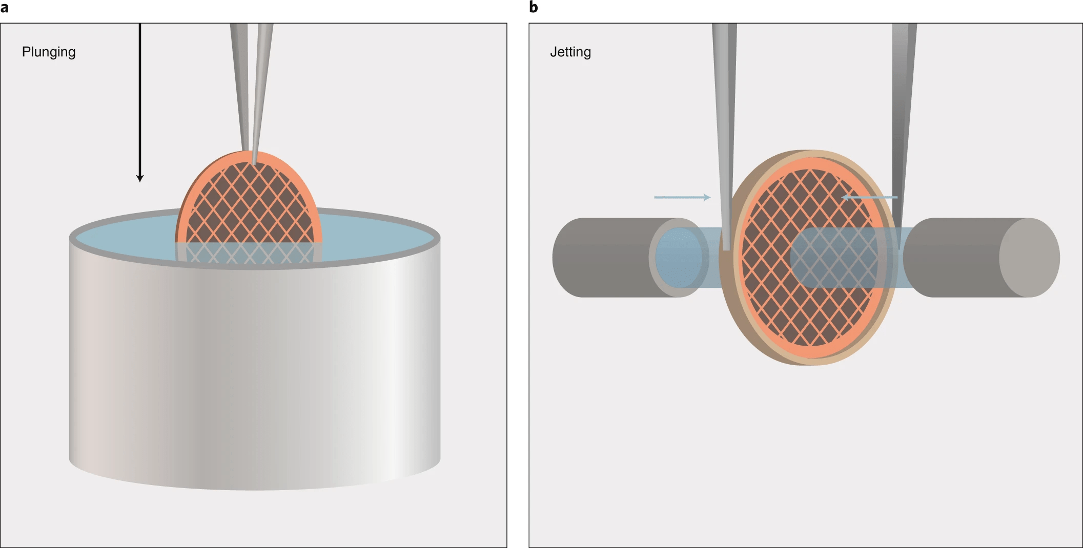
Crystals have the same repeating unit cell, which amplifies our signals
If in solution, particles would be
In Cryo-EM, a beam of high-energy electrons is used instead of photons
Why Electrons?
No crystals: The sample is rapidly frozen in vitreous ice to preserve its native structure
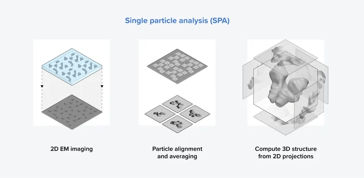
Single Particle Analysis is the main Cryo-EM technique used to determine the 3D structures of individual macromolecules
Molecules are not static
Example: The p53 tumor suppressor protein has flexible regions critical for its regulation and binding interactions
Proteins often exhibit flexibility, disordered regions, and multiple conformations
Why It Matters: Structural techniques often require ordered or stable configurations
One strength of Cryo-EM is its ability to capture multiple conformational states of a molecule, providing insights into flexibility and structural heterogeneity.
Challenge: A major issue in Cryo-EM is that highly flexible or disordered molecules may appear as fuzzy or low-resolution regions in the final structure
Advanced computational techniques are required to sort out different conformations present in the Cryo-EM data
Intrinsically disordered proteins (IDPs) or regions lack a stable 3D structure under physiological conditions but are still functional, often gaining structure upon binding to partners
Lecture 09B:
Structural Biology -
Methodology
Lecture 09A:
Structural Biology -
Foundations
Today
Thursday
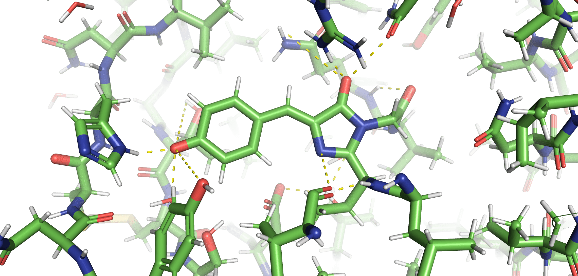
Why is Green Fluorescent Protein (GFP) fluorescent, but not the chromophore in solution?
GFP keeps the chromophore planar and facilitates an excited-state proton transfer
Fluorescent
Not Fluorescent
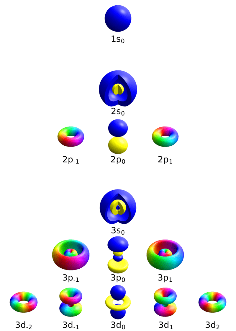
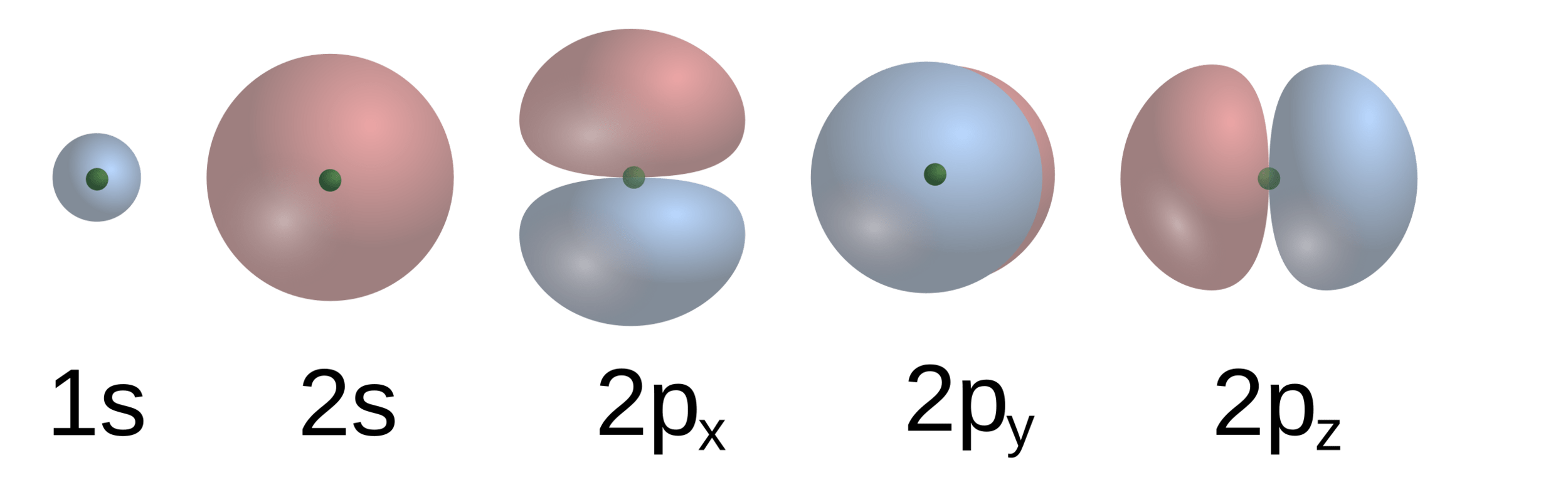
Principle quantum number
1
2
3
Orbital quantum number
0
-1
0
1
-1
0
1
-2
2
Magnetic quantum number
1
1
2
3
2
1
(You don't need to know what these mean)
An electron at (n, l, m) will have a specific energy level and characteristics
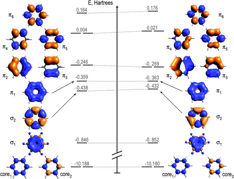
Benzene has . . .
Six carbon atoms with 1s2 2s2 2p2
Six hydrogen atoms with 1s
located at the center of each atom's position
Particles (e.g., electrons and photons) can interact with these molecular orbials
D6h structure
D3h structure
These molecular orbitals determine behavior
Changing the positions (or symmetry) change molecular orbitals
All experimental techniques are based on probes interacting with molecule's electron density to reveal structural information
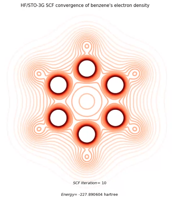
Electron density of benzene
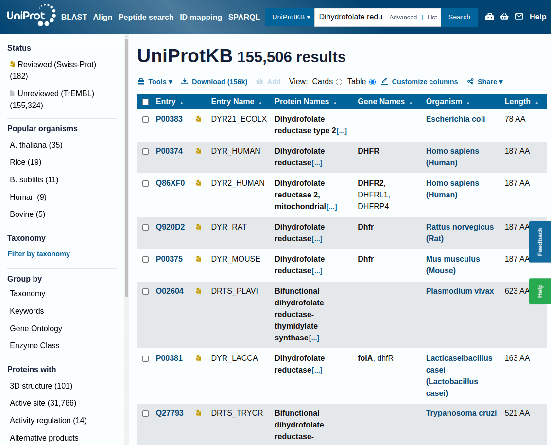
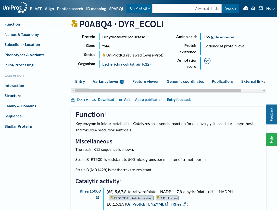
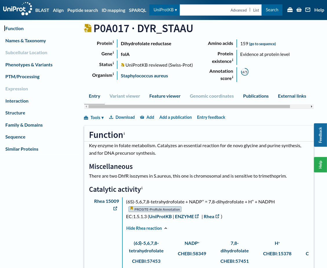
Let's find information about our project's drug target: Dihydrofolate reductase
UniProt is a comprehensive database to access curated data about protein structures, functions, sequences, and annotations.
This page shows the results of a search in UniProtKB for a specific protein, in this case, "Dihydrofolate reductase"
On the left side, you have multiple filters to narrow your search results:
Reviewed (Swiss-Prot): Experts manually curated and verified these entries, ensuring high accuracy
Unreviewed (TrEMBL): These entries are automatically generated and have not been manually reviewed
Each row in the table represents a different protein entry
Entry ID: A unique identifier for the protein (e.g., P00383). You can click on this ID for detailed information about the protein
Many proteins function by switching between different conformations, which is essential for their activity (e.g., enzymes, transporters, and receptors).
Covalent
Covalent bonds are formed when atoms share pairs of electrons that holds molecules together
Strength and stability: Covalent bonds provide the necessary stability for complex biological structures
Directionality: Covalent bonds limit the specific angles and orientations leading to the 3D shapes of biomolecules
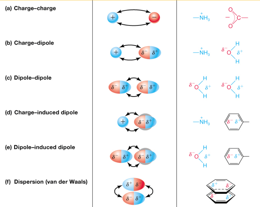
Noncovalent
Noncovalent interactions are weaker than covalent bonds and involve electrostatics
We will cover this in L10
Macromolecular structure
Membrane Formation
Protein-Protein Interactions
Molecular recognition