Dynamic (and static) physiological sources of noise in fMRI data

| smoia | |
| @SteMoia | |
| s.moia.research@gmail.com |
BF-UHF-SG ISMRM Workshop 2025, Annapolis, 01.04.2025
Faculty of Psychology and Neuroscience, Maastricht University, Maastricht, The Netherlands; Open Science Special Interest Group (OHBM); physiopy (https://github.com/physiopy)
I have no financial interests or relationship to disclose with regard to the subject matter of this presentation.
I have the following biases to disclose:
-
I am a member of the Physiopy Community
-
I am a maintainer of the Physiopy packages

Denoising is strongly linked to interpretation
Are you interested in individual or group level effects?
Are physiological responses part of your interpretation?
(e.g. affective neuroscience)
Should your interpretation include autonomic nervous system changes?
[...]
Are your comparisons robust to physiological responses?
Impact of physiology on data variance
1. Bianciardi et al., 2009 (Magn. Reson. Imaging.); 2. Triantafyllou et al., 2005 (NeuroImage);
3. jorge et al., 2013 (Magn. Reson. Imaging.), Reynaud et al., 2017 (Magn. Reson. Med.)
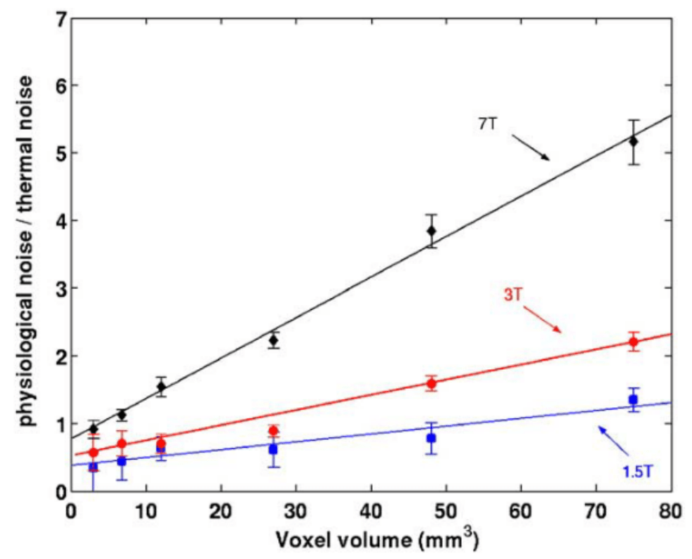
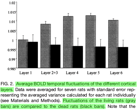
Physiology-related variance varies:
- By voxel size¹ ² and position¹
- By field strength²
- By sequence type and TR³
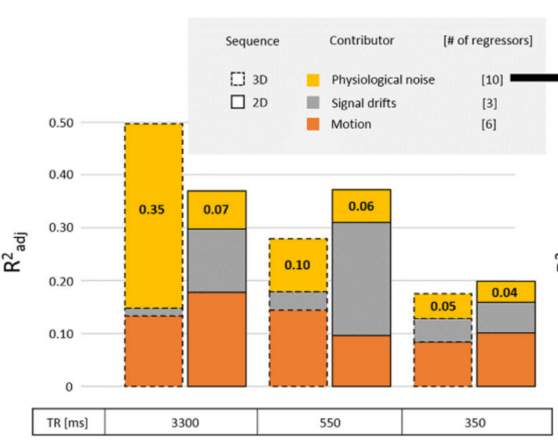
Impact of physiology on data variance
1. Krentz et al., 2023 (bioRxiv), Carlton et al., 2024 (bioRxiv), Moia et al., 2024 (bioRxiv);
2. Birn et al., 2009 (NeuroImage), image courtesy of Jingyuan Chen; 3. Lee et al., 2023 (HBM)
Physiology-related variance varies:
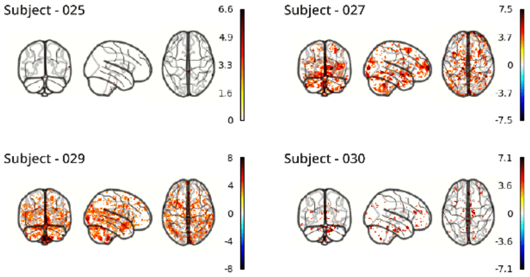
- By individual & session¹
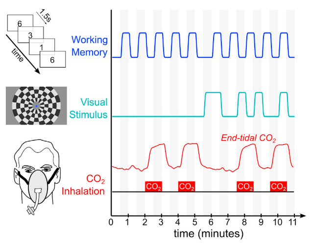
- By task (task-locked)²
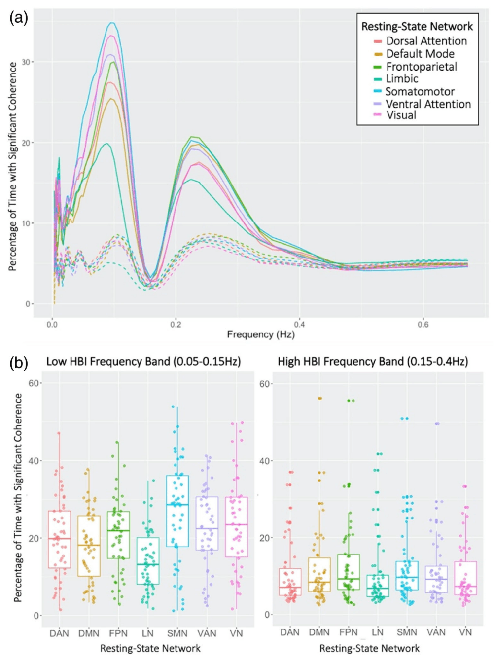
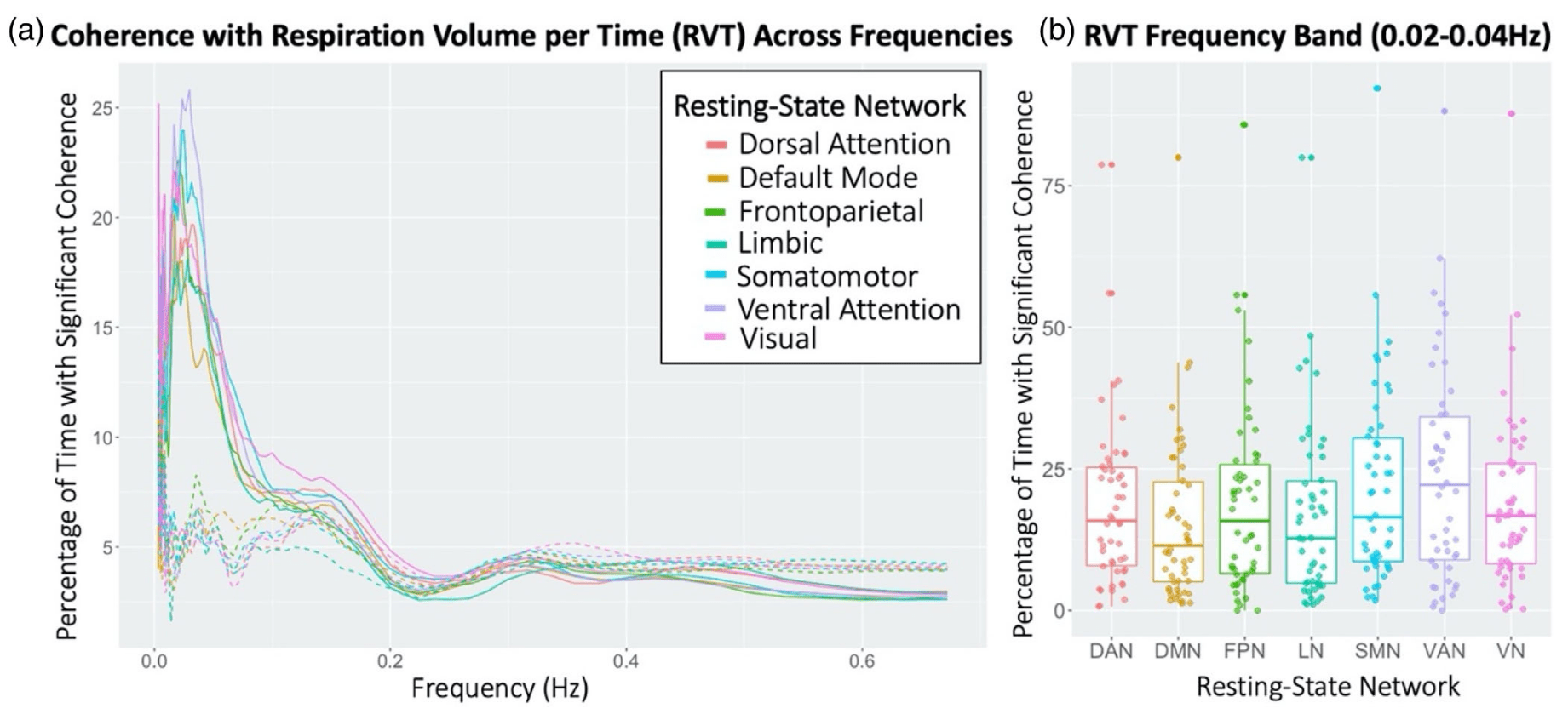
- By Resting State Network³
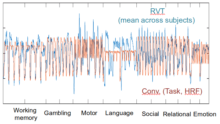
Ventilation
Task (convolved)
RETROICOR variability
How to record physiological data
Ideas on positioning

Ventilation
Respiration (CO2)
Pulse
Electrodermal Activity
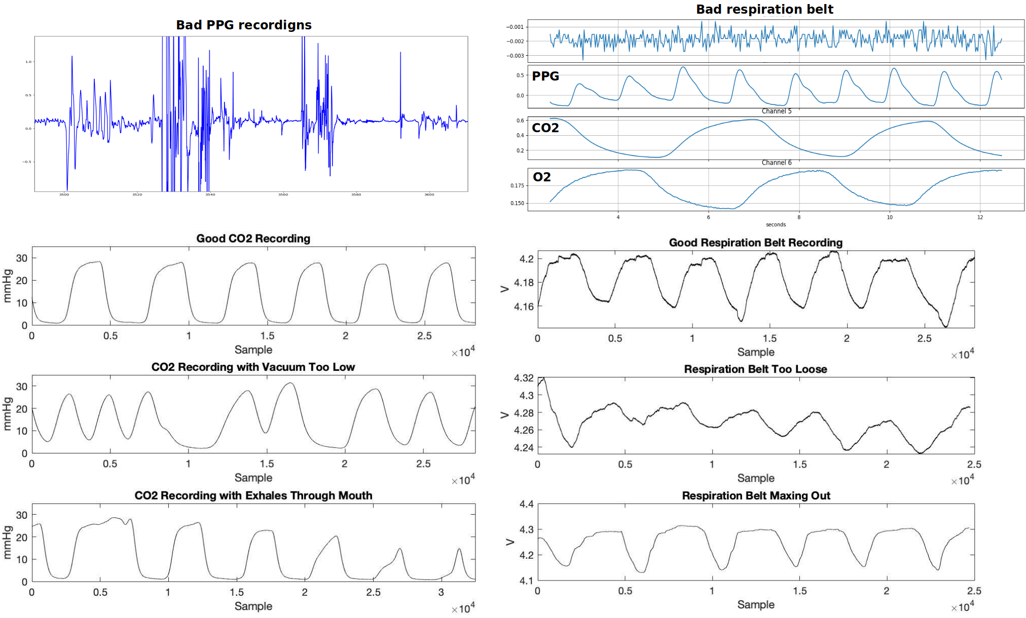
Quality Control
images courtesy of Kristina Zvolanek, Elenor Morgenroth, and César Caballero-Gaudes
"Denoise" the "noise"
Bottenhorn et al., 2023 (Aperture Neuro)
ECG before scanner
ECG during fMRI
ECG after Bottenhorn filter
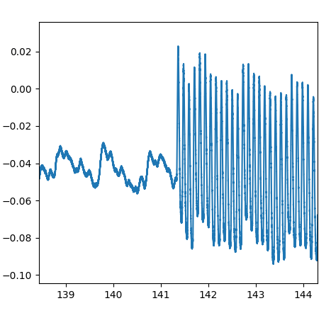
Scanner off
Scanner on
PPG
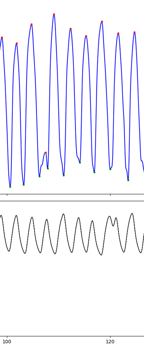
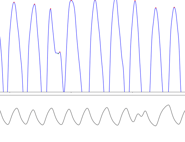
Make sure you're removing what you should
data = np.genfromtxt('sub-007_ses-05_task-rest_run-01_physio.tsv.gz', usecols=[0, 1, 3])
ph = peakdet.Physio(data[:, 1], fs=10000, suppdata=data[:, 2])
ph = peakdet.operations.peakfind_physio(ph, thresh=thr, dist=dist)
ph = peakdet.operations.edit_physio(ph)
DuPre et al., 2024 (Zenodo)
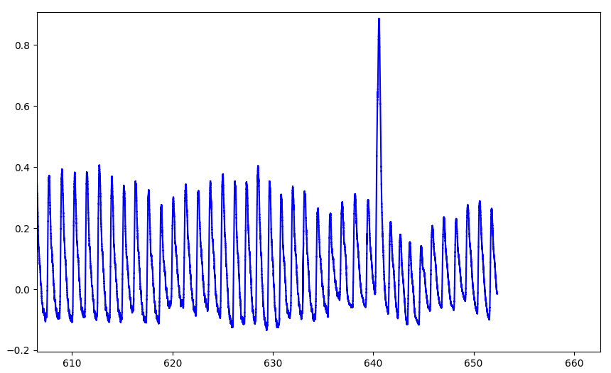
PPG
Ventilation
CO2
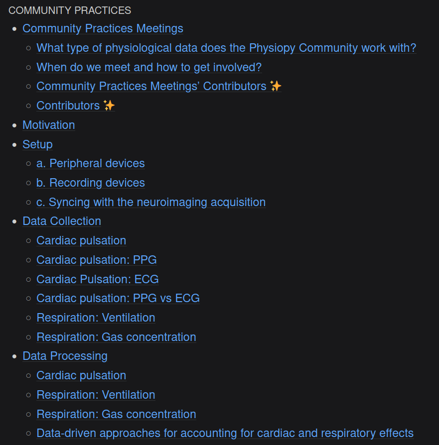
Physiopy Community Practices
physiopy-community-guidelines.readthedocs.io


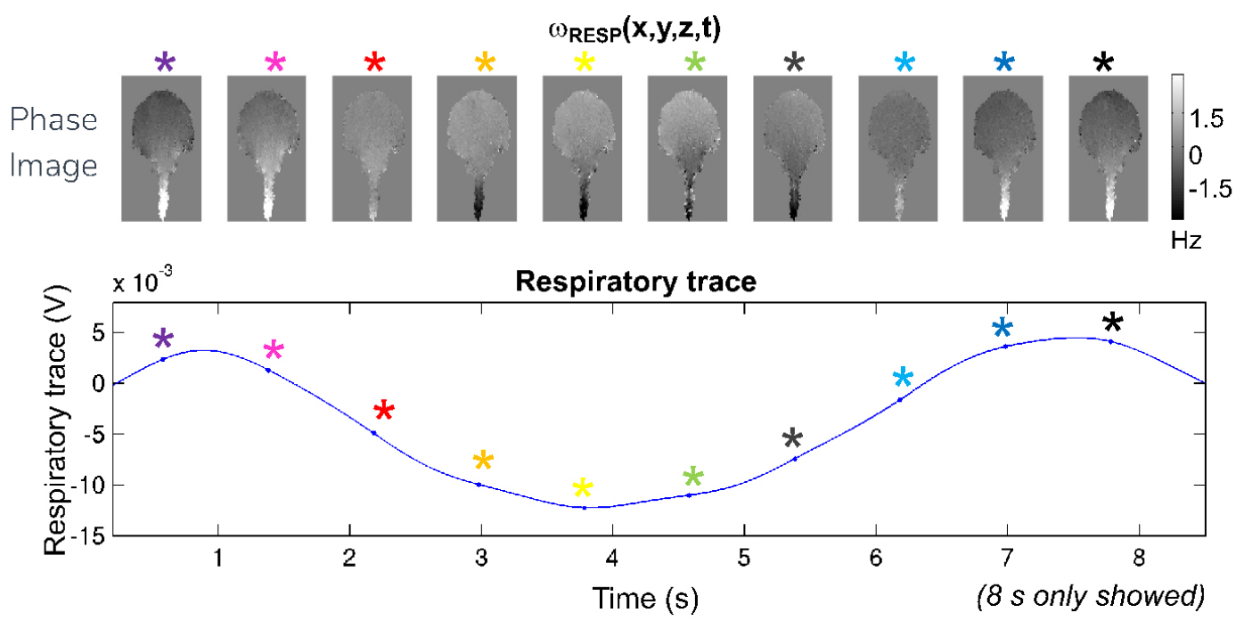
Phase changes over physiological cycles
image courtesy of Marta Bianciardi
Denoising with physiological data: RETROICOR¹
1. Glover et al., 2000 (Magn. Reson. Med.); 2. Kasper et al, 2017 (J. Neurosci. Methods); 3. Krentz et al., 2023 (bioRxiv)
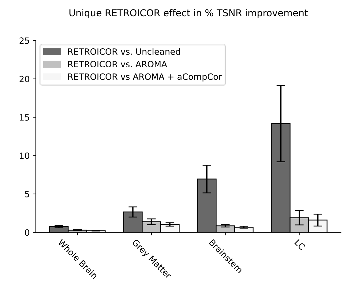
Whole
Brain
GM
Brainstem
LC
RETROICOR variability across subjects³
²
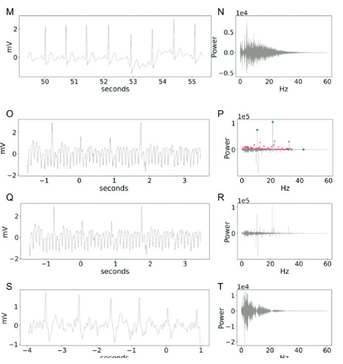
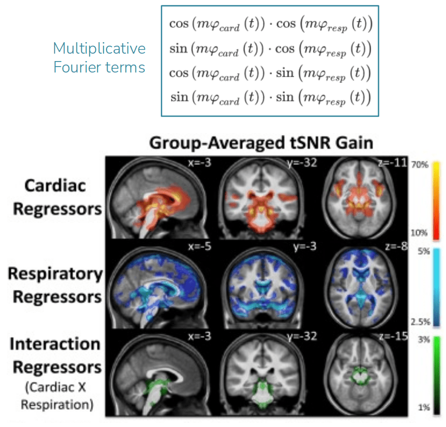
Physiological harmonics
Image courtesy of Blaise Frederick
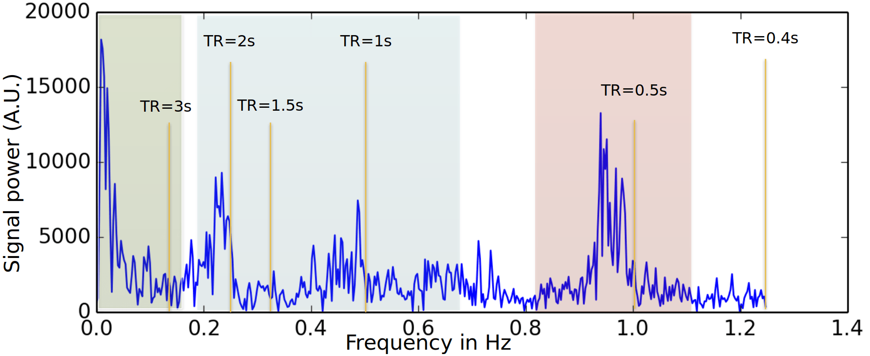
Cardiac
Respiratory
Denoising with physiological data: slow fluctuations
HRV: Shmueli et al., 2007 (NeuroImage); HBI: Chen et al., 2020 (NeuroImage), img from Lee et al., 2023 (HBM)
phys2denoise: Bottenhorn et al., 2024 (Zenodo); phys2cvr: Moia et al., 2024 (Zenodo); PhysIO Toolbox: Kasper et al., 2017 (J. Neurosci. Methods)
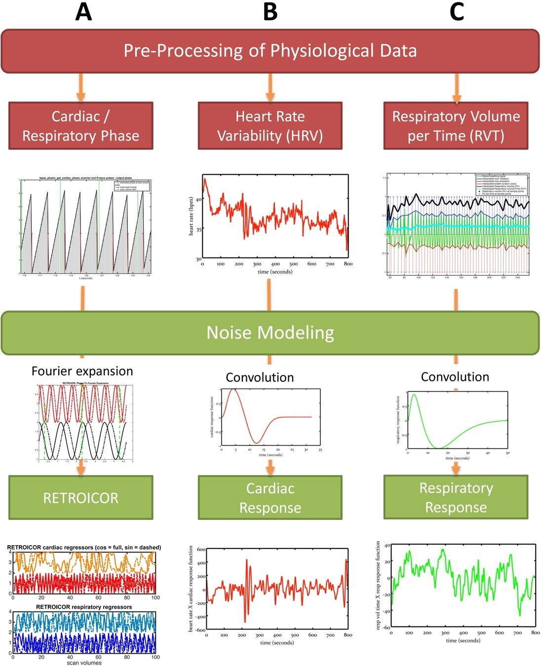
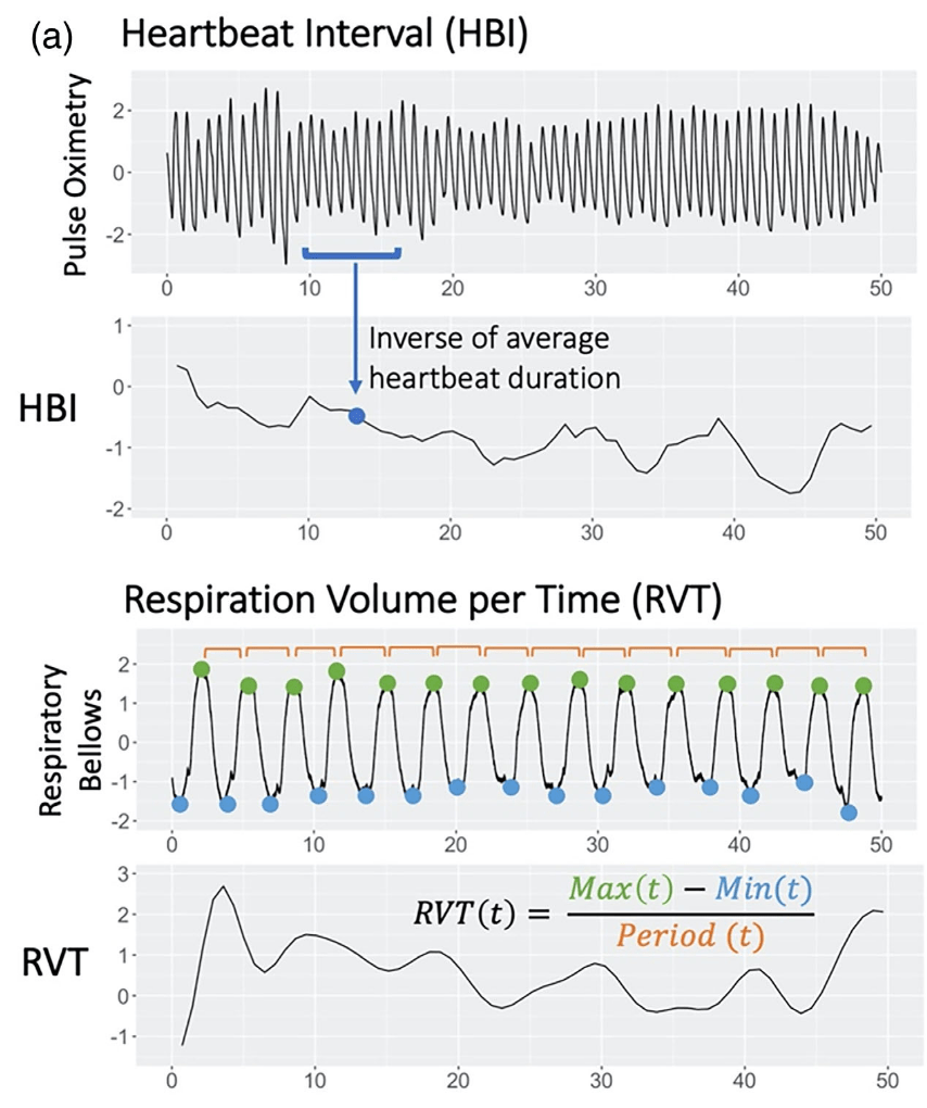
RVT: Birn et al., 2006 (NeuroImage), img from Lee et al., 2023 (HBM); RV: Chang & Glover, 2009 (NeuroImage), img from Chen et al., 2010 (NeuroImage)
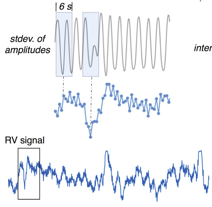
Respiratory Variance (RV)
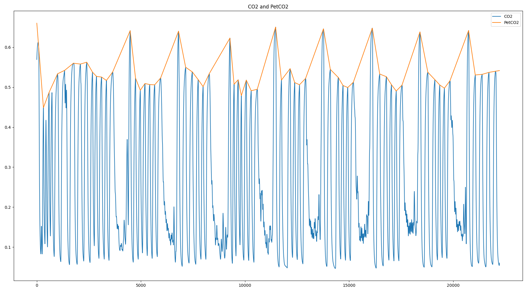
Pressure of End Tidal CO2 (PetCO2)
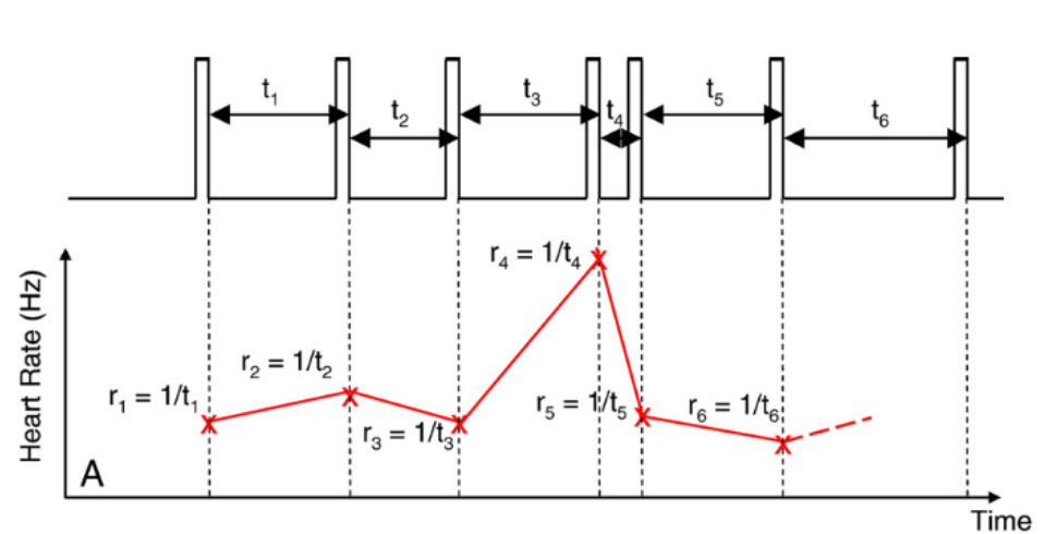
Heart Rate Variability (HRV)
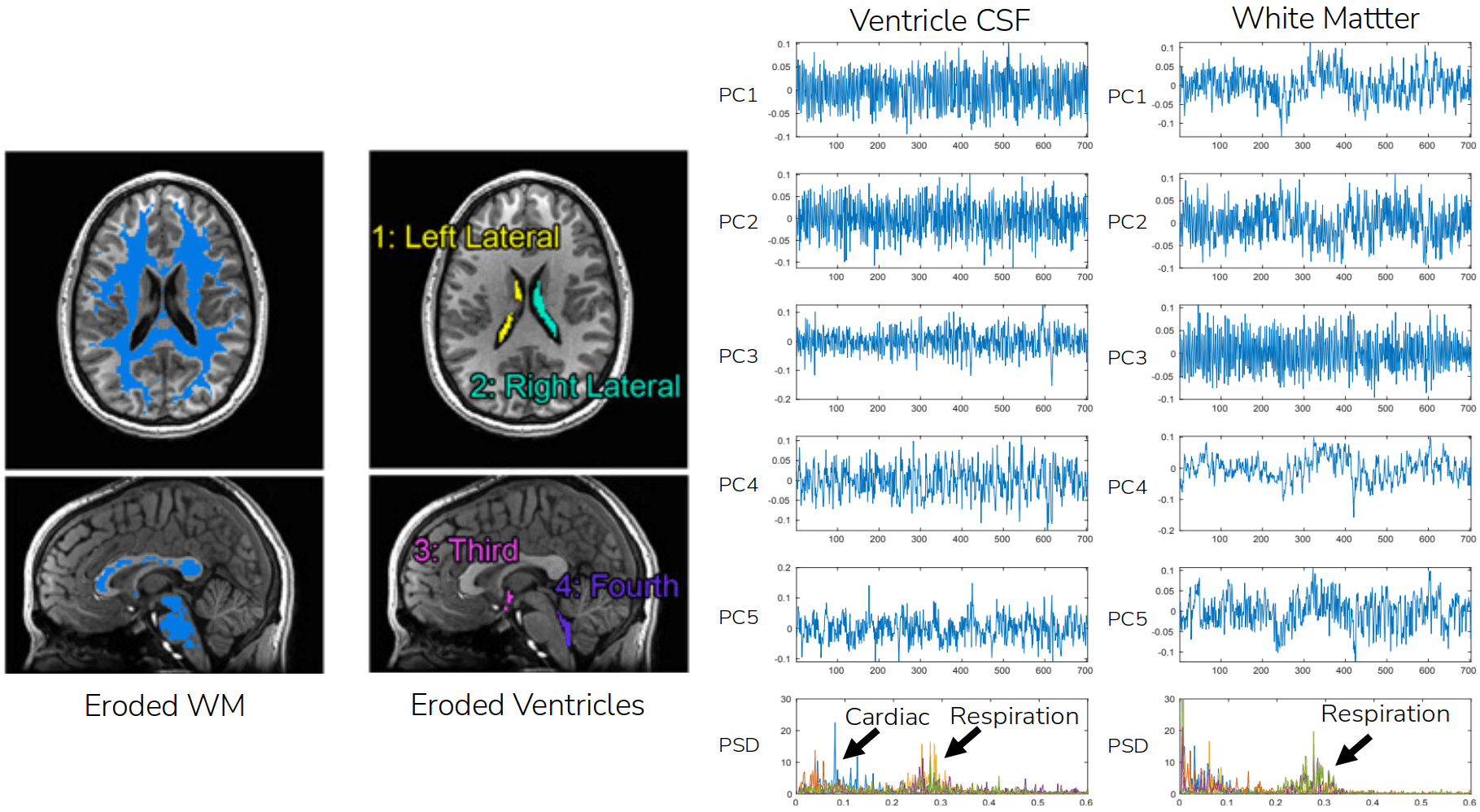
Denoising through data decomposition: CompCorr¹
1. Behzadi et al., 2007 (NeuroImage); image courtesy of César Caballero-Gaudes
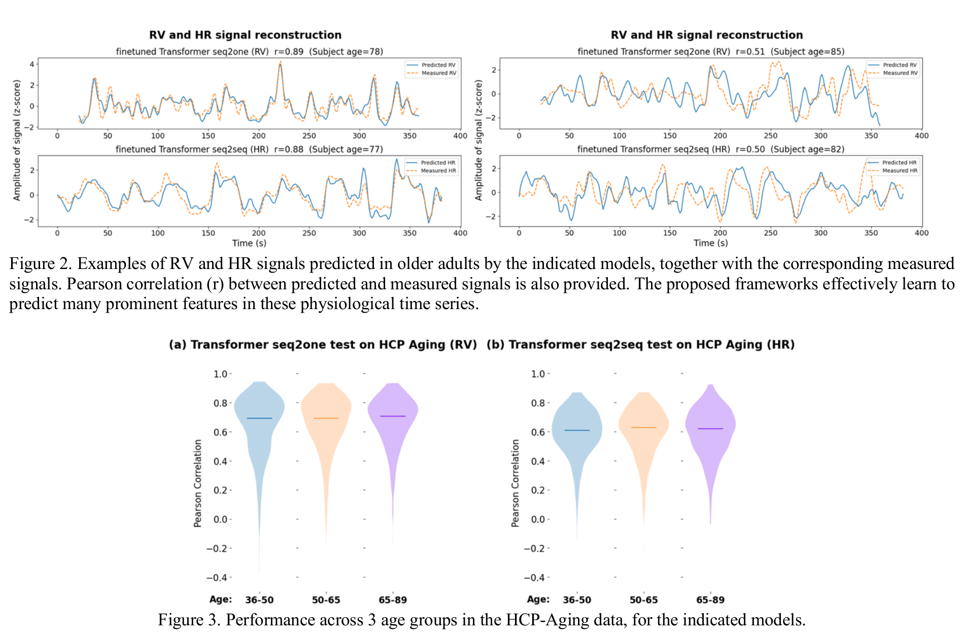
Reconstructing physiological data from fMRI
1. Aslan et al., 2019 (Neuroimage); 2. Wang, Xu, et al., 2024 (arXiv)
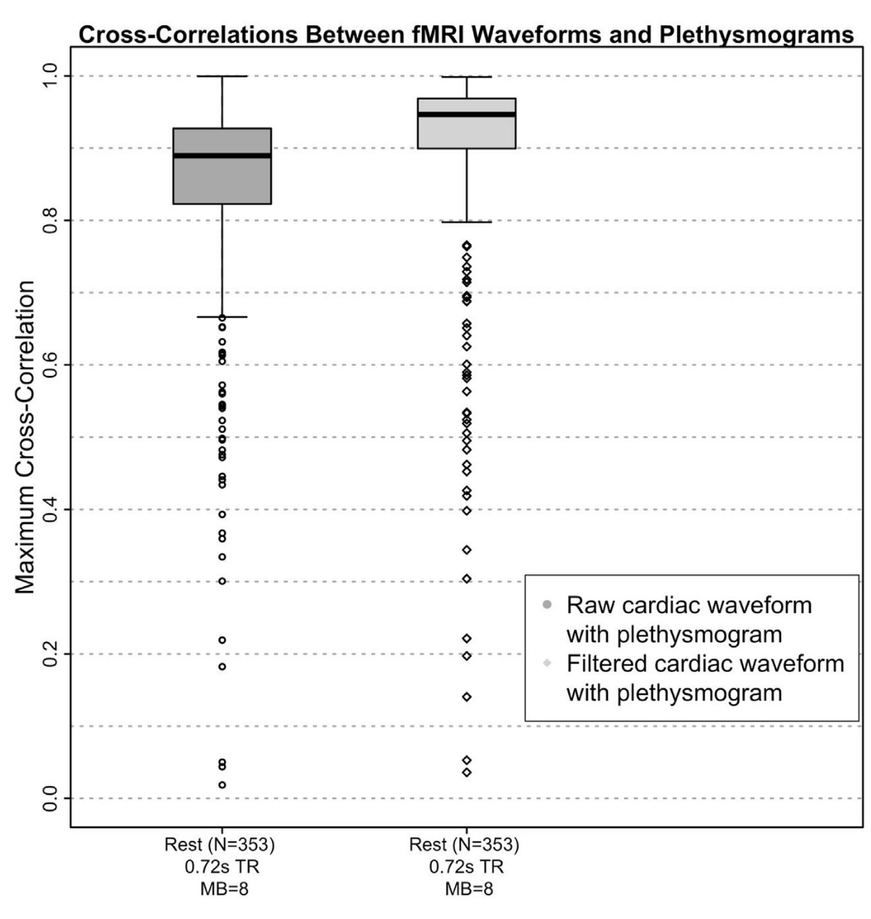
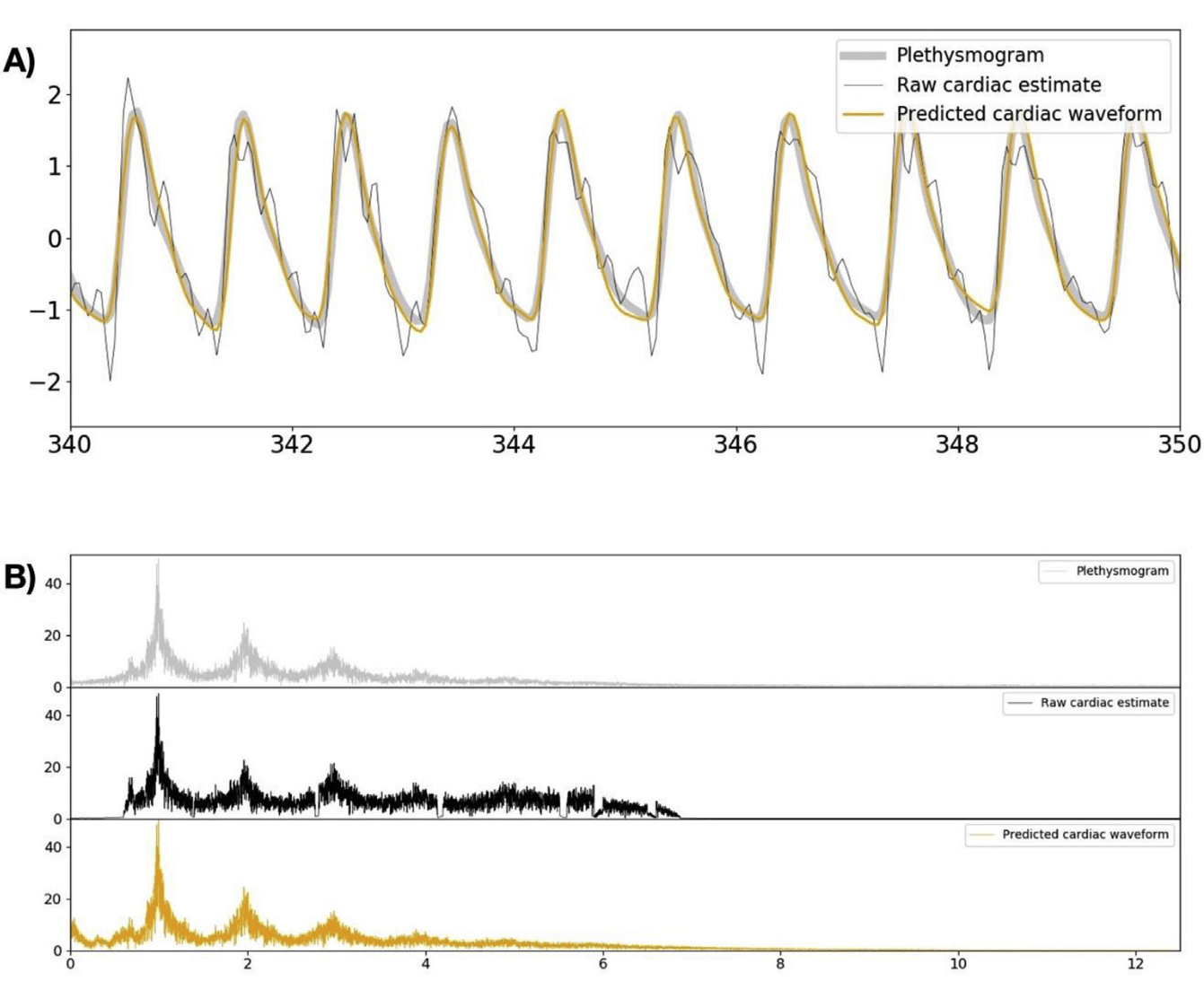
Cardiac waveform from multislice data¹
RV and HRV from parcellated BOLD fMRI²
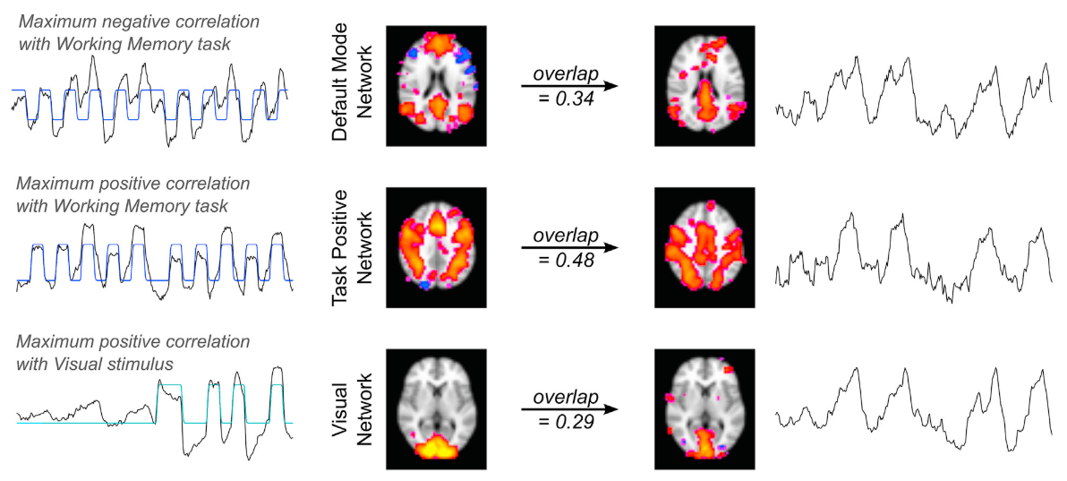
1. Bright et al. 2020 (NeuroImage), Chen et al., 2020 (NeuroImage)
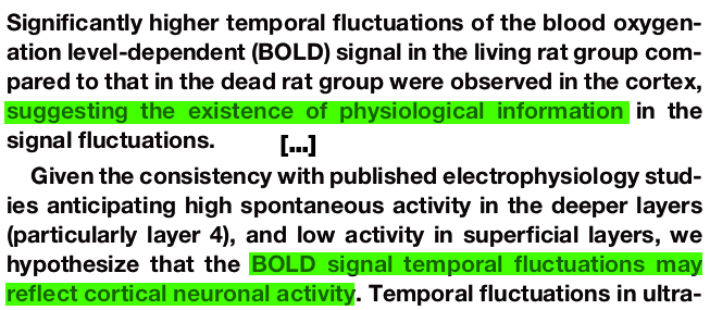
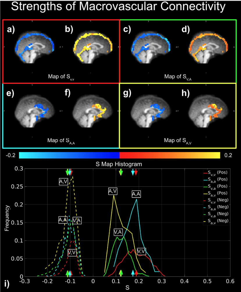
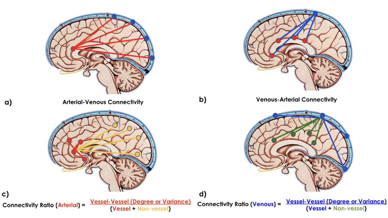
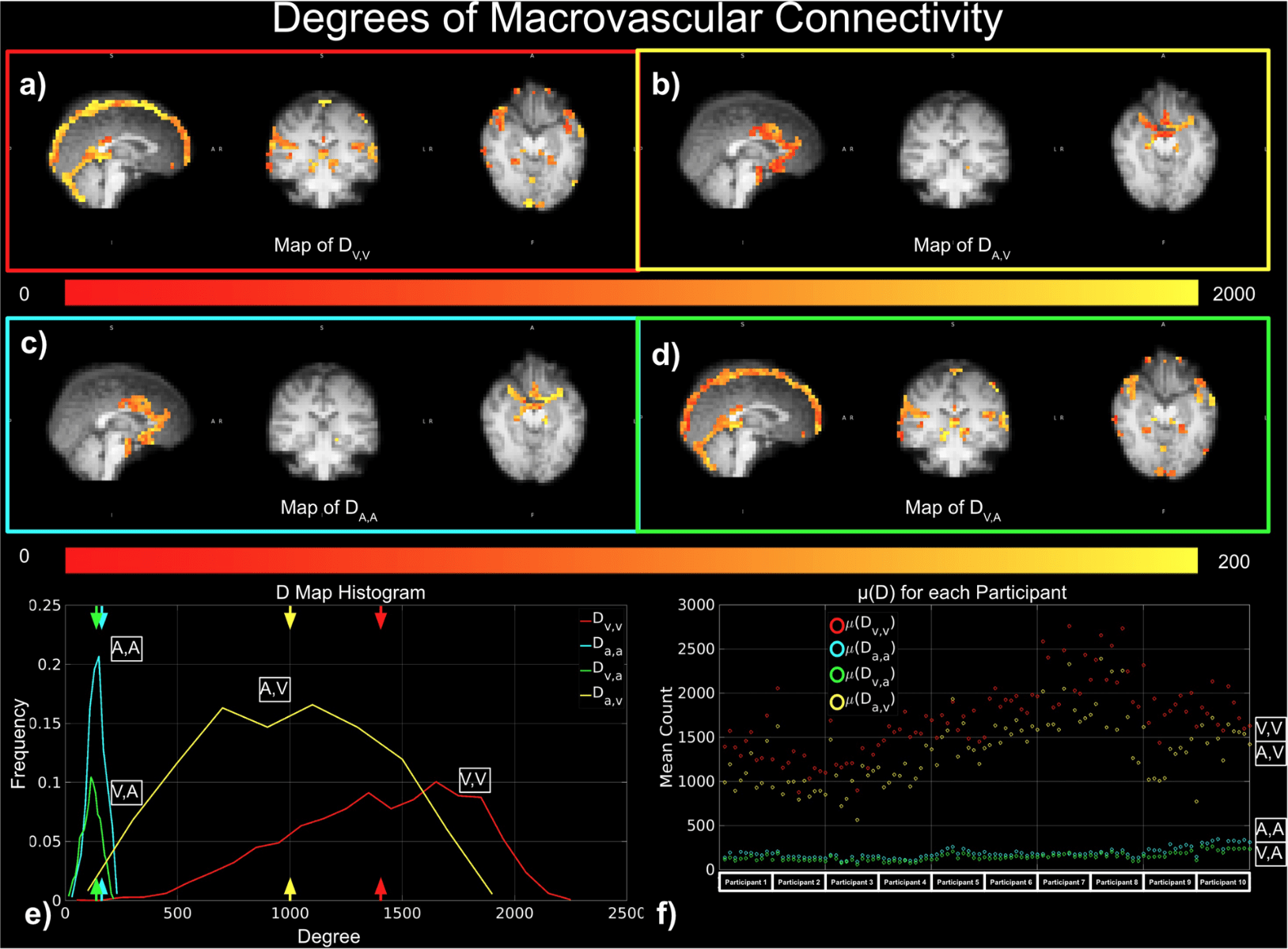
BOLD-based fMRI, in conjunction with physiological signals, reveals the existence of physiological and vascular brain networks¹.
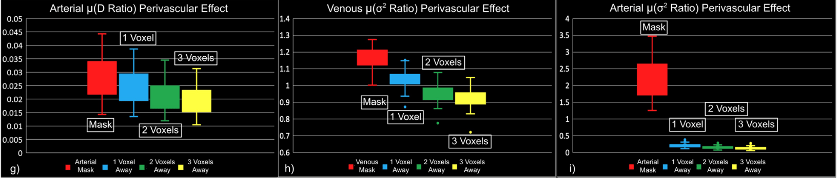
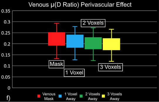
"Static" physiology: vessels
1. Gulban et al., in prep., Gulban et al., 2025 (bioRxiv), image courtesy of Faruk Gulban; 2. Zhong et al., 2024 (Imaging Neurosci.)
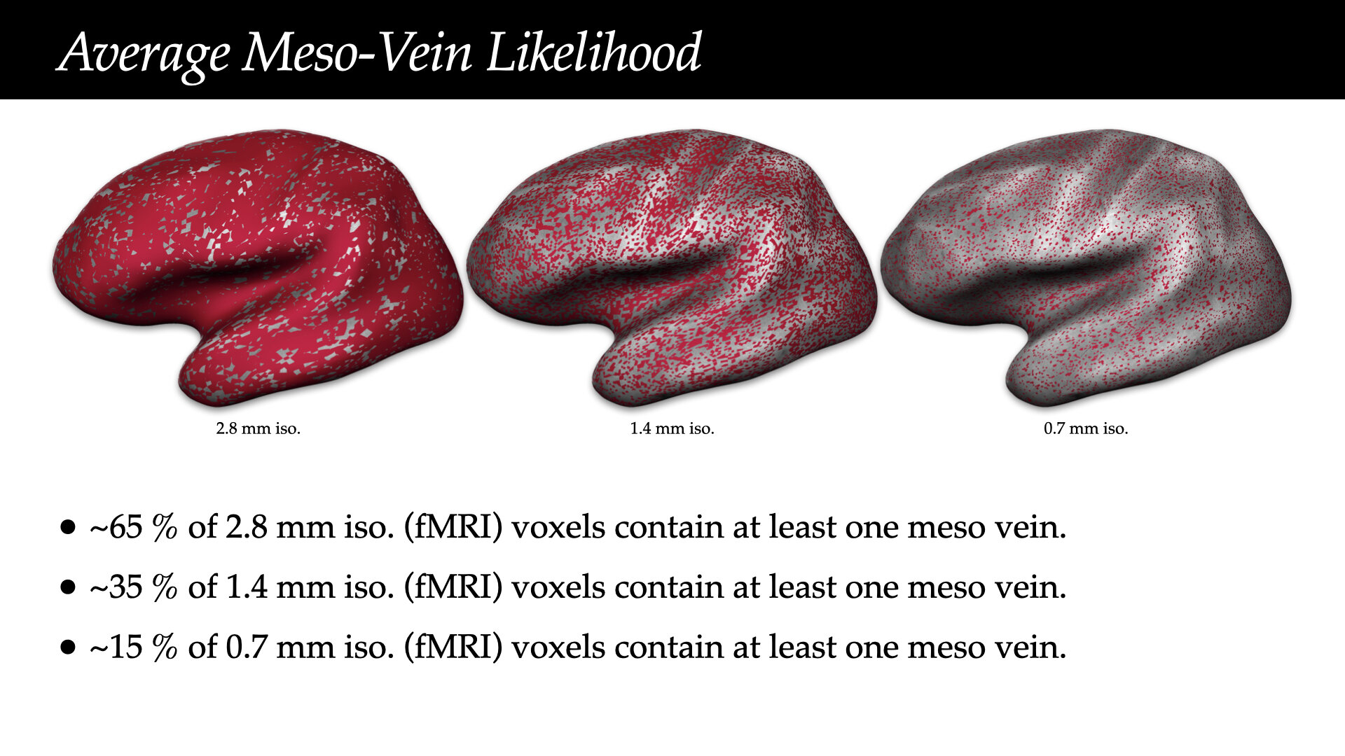
- Meso-scale vessels introduce partial volumes effects
in the grey matter¹ - Macro-scale vessels affect connectivity
in the grey matter²
Denoising is strongly linked to interpretation
"Static" physiology: vessels
Zhong et al., 2024 (Imaging Neurosci.)
That's all folks!
| smoia | |
| @SteMoia | |
| s.moia.research@gmail.com |

Find the presentation at:
slides.com/smoia/
uhf-fmri-2025/scroll


Part of this research supported by the Eunice Kennedy Shriver National Institute of Child Health and Human Development of the National Institutes of Health under award number K12HD073945, the European Union’s Horizon 2020 research and innovation program (Marie Skłodowska-Curie grant agreement No. 713673), a fellowship from La Caixa Foundation (ID 100010434, fellowship code LCF/BQ/IN17/11620063), the Spanish Ministry of Economy and Competitiveness (Ramon y Cajal Fellowship, RYC-2017- 21845), the Spanish State Research Agency (BCBL “Severo Ochoa” excellence accreditation, SEV- 2015-490), the Basque Government (BERC 2018-2021 and PIBA_2019_104), the Spanish Ministry of Science, Innovation and Universities (MICINN; FJCI-2017-31814)


Physiological BIDS
extension proposal(s)

Physiopy Community Guidelines
Any question [/opinions/objections/...]?
Take home messages
- Denoising is linked to interpretation
- Physiological signals should be considered when comparing sequences, subjects, tasks, networks
- Physiological must be quality controlled to avoid bad denoising
- RETROICOR explains part of the data variance that other models do not explain
- When lacking (good) physiological data, data-driven approaches can be used
- Physiological data can be partially retrieved from BOLD MRI
- Brain vessels may bias your functional data
Find the presentation at:
slides.com/smoia/
uhf-fmri-2025/scroll