Week 1 TA Review
Agenda
- Week 1 review: CSF System & Spinal Cord
- Week 2 preview: Brainstem topography and functional levels
- Q&A
Week 1 Review
Ventricular System
On a blank sheet of paper, draw from memory:
- Draw the ventricular system in the sagittal plane.
- Label all ventricles, foramena, and cisterns.
- Draw the direction of CSF flow.
Meninges
On a blank sheet of paper, draw from memory:
- A coronal section of the brain.
- Add a skull
- Draw and label all parts of the meninges.
- Label visible dural folds.
- Which fold(s) is/are not visible in you slice?
- Label all spaces and potential spaces.


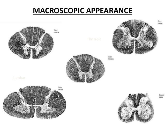
Spinal Cord
On a blank sheet of paper, draw from memory:
- A spinal cord cross section and vertebrae.
- Label:
- Gray matter structures.
- White matter structures.
- Sulci and fissures.
- Nerve roots and ganglions.
- Sections of the meninges.
- Now draw cervical, thoracic, lumbar and sacral spinal cord sections.

Compare your diagrams to class slides
Specimen/fact Quiz
Which answer orders the spinal cord sections from fewest number to greatest number of vertebrae?
- Cervical, thoracic, lumbar, sacral,
- Thoracic, sacral, cervical, lumbar
- Sacral, lumbar, cervical, thoracic
- Lumbar, sacral, cervical, thoracic
Which section has more spinal cord sections than vertebral bodies? How many?
What is the difference between nuclei and ganglia?
- Nuclei are clusters of neuronal bodies in the CNS while, ganglia are clusters of axons in the CNS.
- Nuclei are clusters of axons in the CNS while ganglia are clusters of axons in the CNS
- Nuclei are clusters of neuronal bodies in the PNS while ganglia are clusters of neuronal bodies in the CNS.
- Nuclei are clusters of neuronal bodies in the CNS while ganglia are clusters of neuronal bodies in the PNS.
At what vertebral level is the highest at which a lumbar puncture is generally safe?
- L2/L3 interspace
- L3/L4 interspace
- L4/L5 interspace
- L5/S1 interspace
Why?
(What structure are we avoiding?)
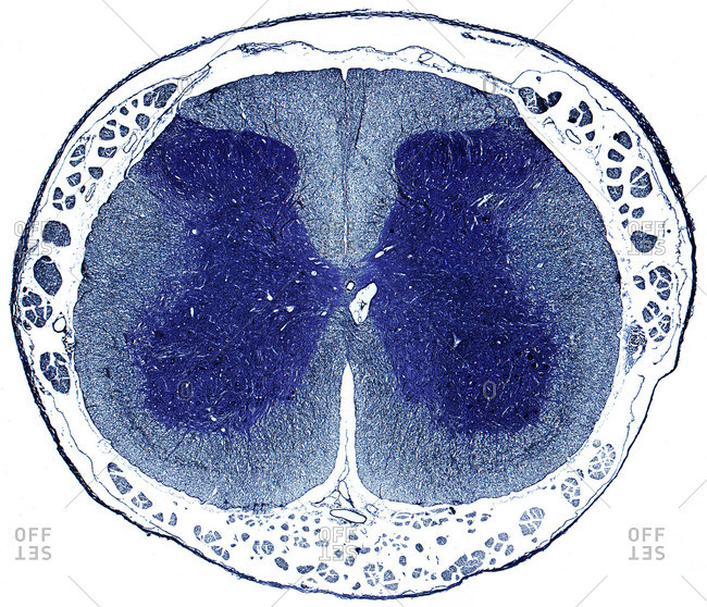
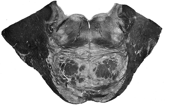
Name that section

Point to the:
- Ventral horn
- Dorsal horn
- Lateral horn
- Intermediate zone
- Anterior funiculus
- Lateral funiculus
- Posterior funiculus
- Anterior median fissure
- Posterior median sulcus
- Anterolateral sulcus
- Posterolateral sulcus
- Ventral root
- Dorsal root
- Dorsal root ganglion
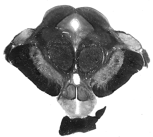
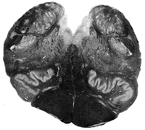
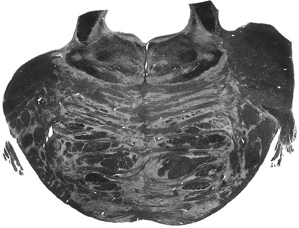
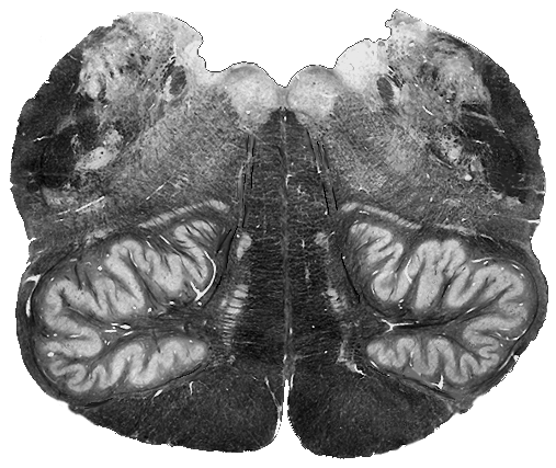
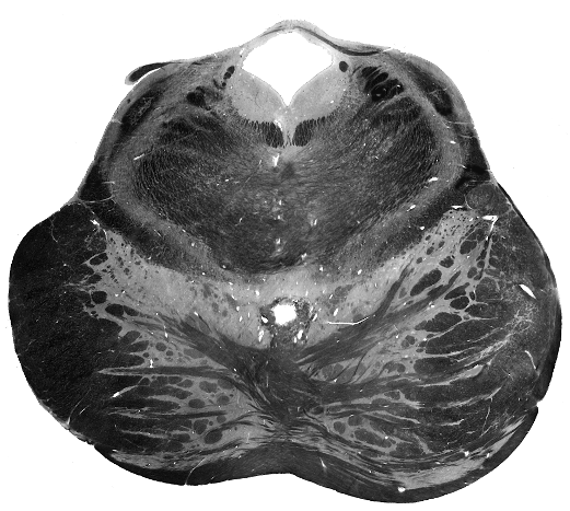
Name that section
Point to the:
- Ventral horn
- Dorsal horn
- Lateral horn
- Intermediate zone
- Anterior funiculus
- Lateral funiculus
- Posterior funiculus
- Anterior median fissure
- Posterior median sulcus
- Anterolateral sulcus
- Posterolateral sulcus
- Ventral root
- Dorsal root
- Dorsal root ganglion
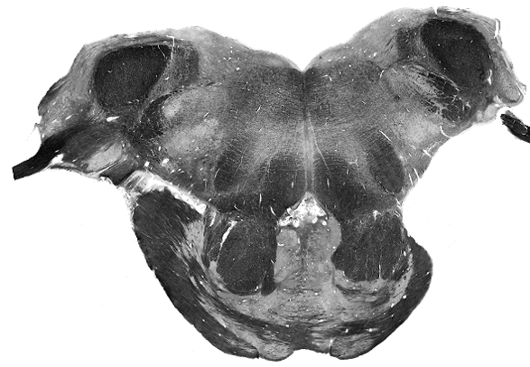
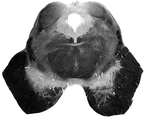
Name that section
Point to the:
- Ventral horn
- Dorsal horn
- Lateral horn
- Intermediate zone
- Anterior funiculus
- Lateral funiculus
- Posterior funiculus
- Anterior median fissure
- Posterior median sulcus
- Anterolateral sulcus
- Posterolateral sulcus
- Ventral root
- Dorsal root
- Dorsal root ganglion
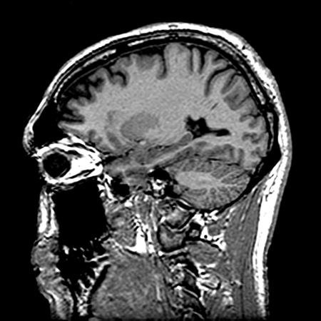
Compare
What ventricles can you see?

Name the structure
Name the structure
Name the structure

Name the structure

Name the structure
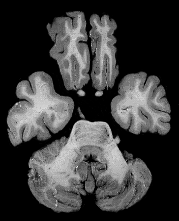
Name the structure
Week 2 Preview
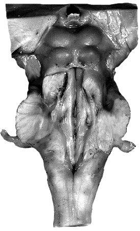
Anterior Brainstem
Anterior Brainstem
What should be here?
What should be here?
Anterior Brainstem
Anterior Brainstem
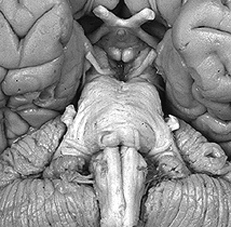
Posterior Brainstem
Caudal Medulla
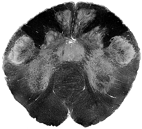
Caudal/Rostral Medulla
Rostral Medulla
Rostral Medulla
Caudal Pons
Middle Pons
Rostral Pons
Caudal Midbrain
Rostral Midbrain