Week 4 TA Review
Outline
- Week 5 preview: Lower Motor Neurons - Cranial and Spinal Cord
- Week 4 review: Forebrain Topography and Functional Levels and the Basal Ganglia
- Q&A
Week 5 Preview
Activity
Grab a brain model. Find the cranial nerves on the model. Point to them. State their function, signs and laterality (ipsilateral, bilateral and contralateral).
Navigate to Washington brain atlas sections. Can you find the nerves and their nuclei?
Fill in the box
| Nerve/Nucleus | Function | Signs (I/B/C) |
|---|---|---|
| Oculomotor (CN III) | Superior levator, superior rectus, medial rectus, inferior rectus and inferior oblique eye muscles | Ipsilateral Ptosis (drooping eye lid) Mydriasis (dilated pupil) Opthalmoplegia (paralysis of eye muscles) Down & out eye |
| Trochlear (CN IV) | Superior oblique eye muscle | Contralateral to nuclues Ipsilateral to nerve Extorsion (hard to look down and in) Unconscious head tilt |
| Trigeminal (CN V) | Muscles of mastication Lateral pterygoid Medial pterygoid Masseter |
Ipsilateral Muscular atrophy Jaw deviation towards side of lesion Difficulty chewing |
| Abducens (CN VI) | Lateral rectus muscle | Ipsilateral Esotropia (cannot abduct eye) |
| Facial (CN VII) | Muscles of facial expression | Ipsilateral Paralysis of facial expression muscles Lack of salivation, lacrimation Hyperacusis |
| Glossopharyngeal (CN IX), Vagus (CN X), Accessrory (CN XI) | Palatal, pharyngeal, and laryngeal muscles, accesory spinal muscles | Ipsilateral Vocal muscle paralysis Dysphagia Contralateral deivation of uvula |
| Hypoglossal (CN XII) | Tongue muscles | Ipsilateral Atrophy Ipsilateral deviation of the tongue |
Define lower motor neuron syndrome.
What is the mechanism of these symptoms?
Specimens
Give the name, function and symptoms of the following structures.
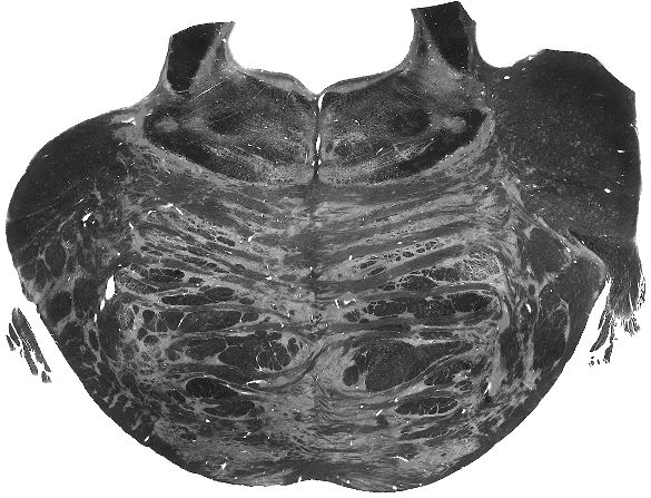
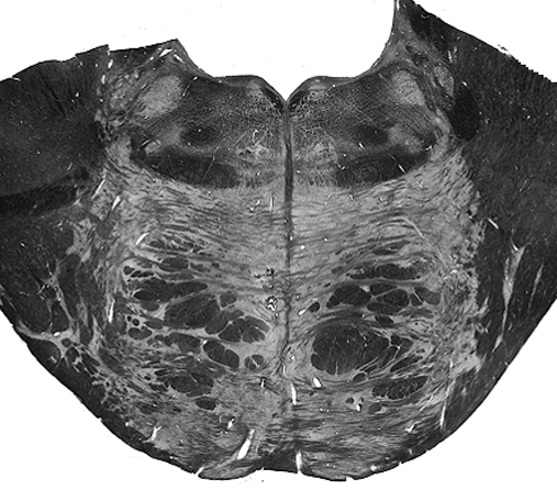
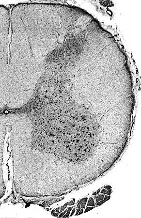
LMN Spinal injury
This section came from L2 of the spinal cord.
What symptoms generally would you expect to see if there was a lesion in this region of the spinal cord?
Myotatic Reflex

Draw a diagram of the Myotactic reflex arc.
Inverse Myotatic Reflex

Draw a diagram of the inverse myotactic reflex arc (Golgi tendon reflex).
Week 4 Review
Warm-up Exercises
Atlas Cruise
In BrainStorm, select gross dissections. Starting in the dorsal orientation, find the following structures:
Can you do it in ventral, midsagittal or sagittal orientations?
- Superior frontal gyrus
- Superior frontal sulcus
- Middle frontal gyrus
- Inferior frontal sulcus
- Inferior frontal gyus
- Lateral sulcus/fissure
- Precentral sulcus
- Precentral gyrus
- Central sulcus
- Postcentral gyrus
- Postcentral sulcus
- Superior parietal lobule
- Intraparietal sulcus
- Inferior parietal lobule
- Anterior paracentral lobule
- Posterior paracentral lobule
- Marginal sulcus
- Precuneus
- Supramarginal gyrus
- Angular gyrus
- Occipital gyri
- Superior temporal gyrus
- Superior temporal sulcus
- Middle temporal gyrus
- Inferior temporal sulcus
- Inferior temporal gyrus
- Cingulate gyrus
- Corpus callosum
- rostrum
- genu
- body
- splenium
- Parieto-occipital sulcus
- Cuneus
- Calcarine sulcus
- Lingual gyrus
- Uncus
- Occipitotemporal gyrus
- Parahippocampal gyrus
- Longitudinal fissure
- Collateral sulcus
- Occipitotemporal sulcus
- Preoccipital notch
Fact Quiz
Instructions:
The following questions are multiple choice. The answer options will be hidden at first. If possible, try to answer the question without seeing the options first.
- Internal capsule
- Corpus callosum
- Anterior commisure
- Corona radiata
What is the primary projection fiber system of the brain?
- Contralateral homonymous hemianopsia (on the right side)
- Contralateral homonymous hemianopsia (on the left side)
- Contralateral neglect syndrome (on the left side)
- Contralateral neglect syndrome (on the right side)
An individual has sustained a lesion to their right calcarine sulcus. What deficits should be present?
- the premotor cortex, paresis
- the parietal association cortex, astereognosis
- the primary visual cortex, visual agnosia
- the temporal association cortex, inability to localize sound
A lesion within _______ is characterized by ______ .
- Cingulate sulcus, cingulate gyrus.
- Inferior temporal sulcus, inferior temporal gyrus.
- Collateral sulcus, parahippocampal gyrus.
- Marginal sulcus, precuneus.
________ borders the fusiform gyrus and the _______ .
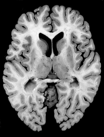
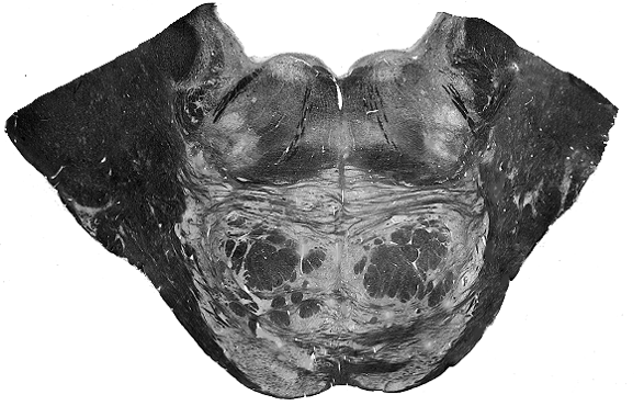
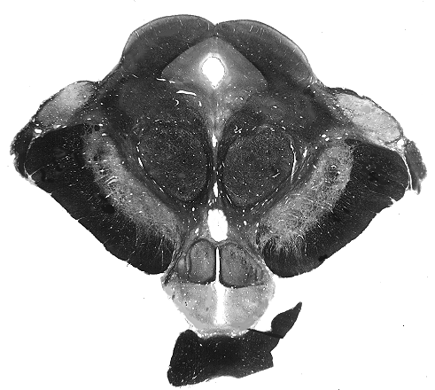
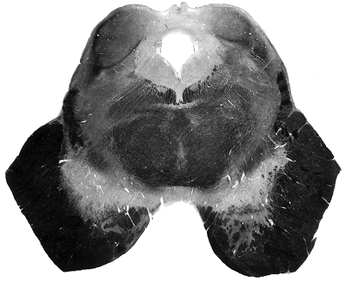
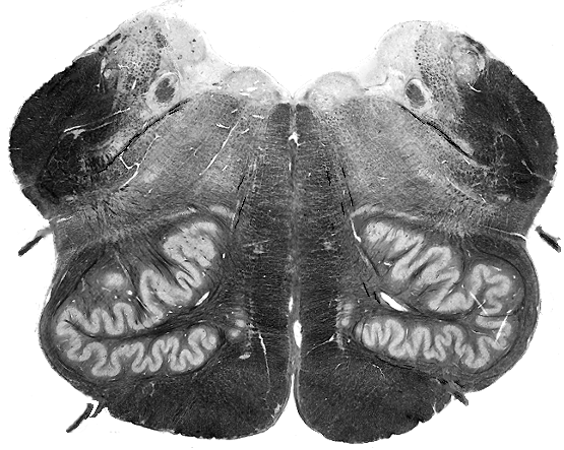
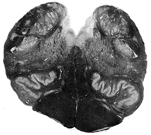
Specimen/Image Quiz
Label the structure
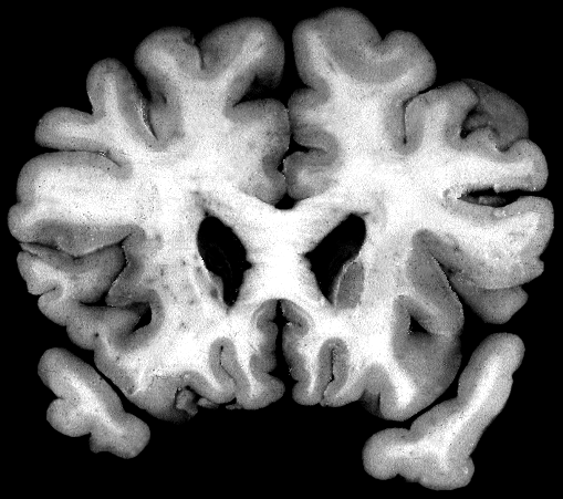
Label the structure
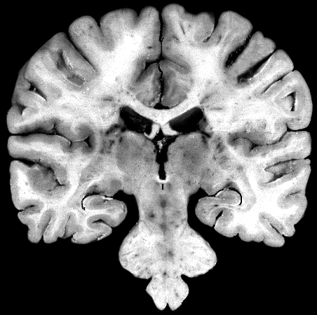
Label the structure
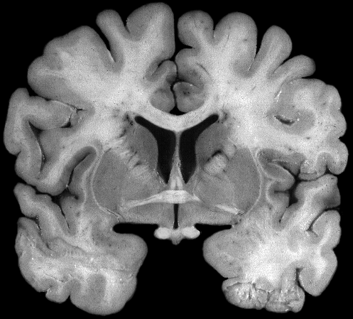
Label the structure