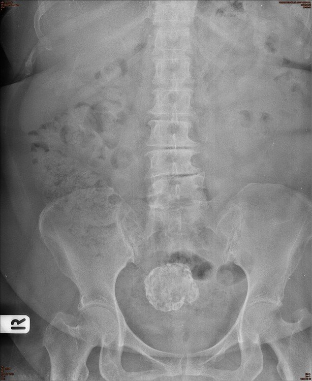
Abdomen : Case 5
History : A 52-year-old male with abdominal discomfort
What is the abnormality?
A. Bladder stone
B. Calcified uterine myoma
C. Calcified node
D. Artifact
Answer : B . Calcified uterine myoma or calcified fibroid
Uterine Myoma:
- Benign tumors of smooth muscle origin
- 20-50% of women > 30 years of age
- Fibroids may enlarge with elevated estrogen levels such as during the first trimester of pregnancy
- Uterine fibroids diminish in size after menopause
Clinical Findings :
- Most : Asymptomatic
- Symptoms can include : Menorrhagia, pain, urinary frequency, urgency, incontinence or Infertility
Imaging Findings : Ultrasound is the study of choice
-
Ultrasound
- Echogenic mass if fibrosis prevails
- Hypoechoic, solid mass if muscle component is prevalent
- Sharp discrete shadows
- Anechoic features if central portion of fibroid has degenerated
-
Conventional radiography :
- Soft tissue mass arising from the pelvis but separate from the urinary bladder
-
Amorphous, flocculent calcifications in the pelvis
- May resemble “popcorn” or may calcify the outer rim of fibroid
- Displacement of bowel gas up and out of the pelvis
Complications :
- Rarely (< 1%), malignant degeneration to a leiomyosarcoma
- Exophytic fibroid can torse and cause pain
- Infertility from interference with implantation
- During pregnancy< >Spontaneous abortionIntrauterine growth retardation
- Postpartum hemorrhage
