Medical Imaging
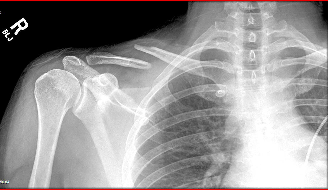
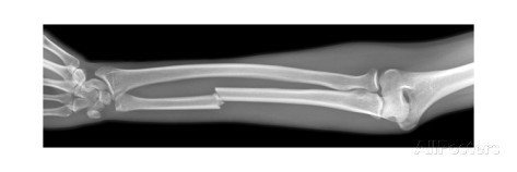
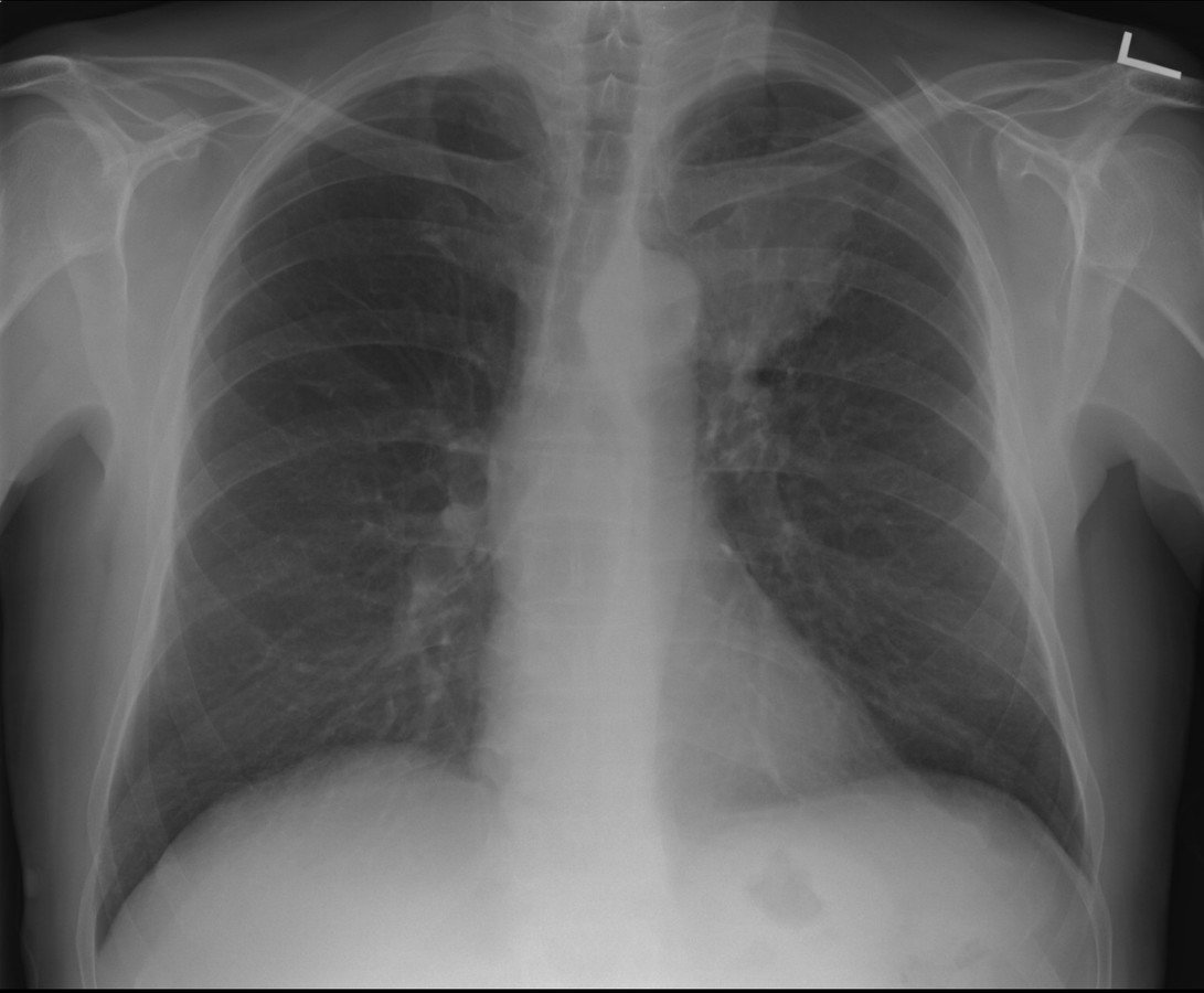
Radiography
(X-Rays)
- Uses ionizing radiation
-
How does it work?
- X-ray beam passed through the body
- Portion of the beam is scattered or absorbed by bones, organs, etc.
- Remaining pattern is transmitted to a detector for further processing as a picture
- Useful for detecting bone problems, infection, and tumors
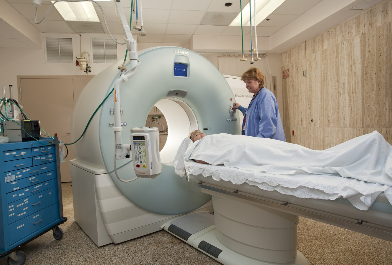
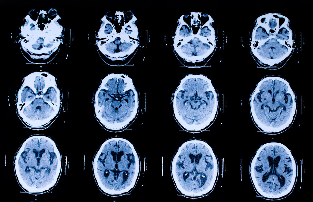
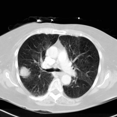
Computed Tomography Scan
(CT Scan)
- More powerful ionizing radiation than X-ray
-
Takes a 360-degree image of internal organs, spine, and vertebrae
- Gives pictures in cross-sections
- More detailed look at organs, soft tissue, blood vessels
- Better diagnostic for cancer, heart disease, infection
- More expensive than X-ray
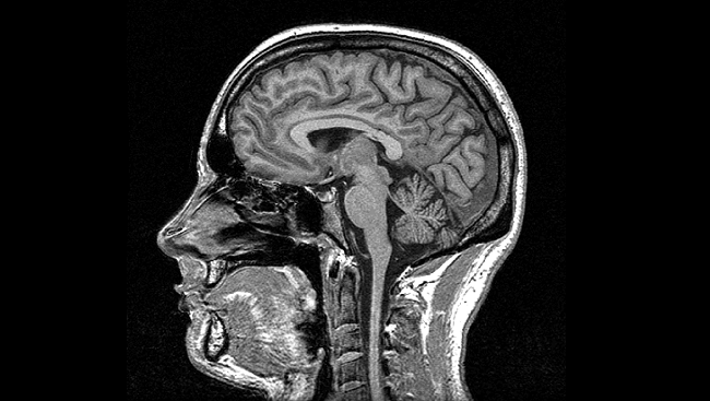
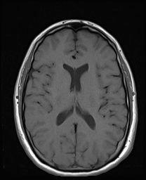
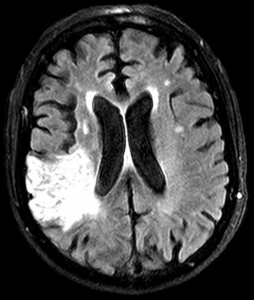
Magnetic Resonance Imaging
(MRI)
- Uses a magnetic field and pulses of radio wave energy to make pictures
- Used to detect tumors, bleeding, injury, blood vessel disease, or infection
- The best tool to detect a stroke
- Most expensive imaging study
- Must remove all metal prior to exam
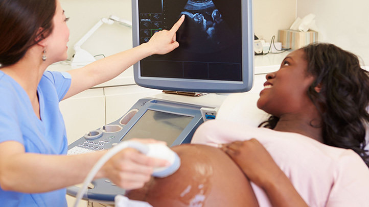
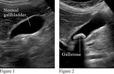
Ultrasound Scan
- Uses high frequency sound waves to create images from inside the body
- Sound waves travel through soft tissue and fluids but bounce off of denser tissue
- The returning sound waves (echo) are measured and used to produce a picture
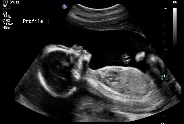
- Does not use radiation--preferred method for pregnant women
- Often used for imaging internal organs found in the abdomen (ex. gallbladder, liver)
- Used to see babies in utero
Medical Imaging
By szondor