BIOPSCHOLOGY
SPEC
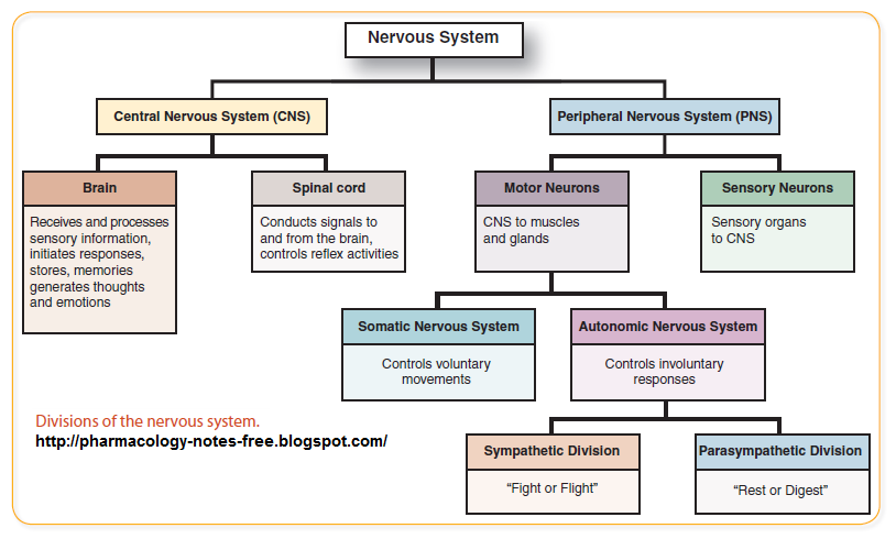
- Divisions of the nervous system
- The different types of neurons
- Synaptic transmission
- Endocrine system
- Fight or flight response
PARTS OF THE BRAIN
| Part | Functiooonnnnn |
|---|---|
| Cerebellum | MOTOR SKILLS AND BALANCE |
| Parietal | SENSORY INFORMATION |
| Temporal | MEMORY AND HEARING |
| Frontal | HIGHER/COMPLEX THINKING |
| Occipital | VISION + SEEING |
Cerebrum= largest part of the brain. Divided into two hemispheres, into four lobes.
Cerebellum- not that. a lobe. controls motor skills and balance
Spinal Chord
relays info between your muscles/body and your brain.
basically an extension of the brain.
Connects nerves to peripheral nervous system.
Peripheral Nervous System!
Autonomic= Involuntary movements such as breathing, heart beat, digestion, stress response . Carries commands from the brain stem.
Somatic= Voluntary muscle movement. Transmits info from the sensory receptors to CNS. Carries commands from the motor cortex. Only excitatory movement.
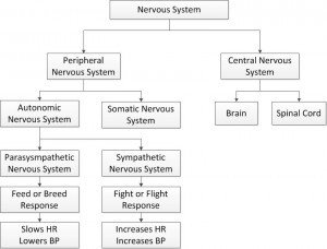
Autonomic: Sympathetic and Parasympathetic NS
| Sympa | Parasympa |
|---|---|
| Gets your body excited af | Chills your body out |
| Prepares your body for fight or flight response | Known as the rest and digest system |
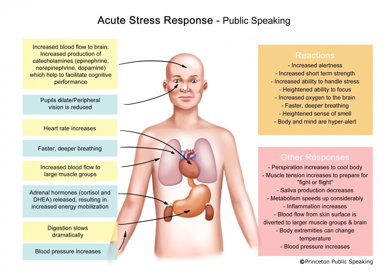
FIGHT OR FLIGHT RESPONSE
Acute response:
When our body sees a stressor, the AMYGDALA associates what we see with an emotion. The stressor sends a signal to the hypothalamus.
The hypothalamus sends nerve impulses along the SYMPA to prepare the body for immediate acions. It goes in several places, one is the adrenal medulla.
SO your adrenal medulla (in the pancreas) secretes adrenaline and nor-adrenaline into the bloodstream. This amplifies the effects of the normal sympathetic nervous system.
Your pupils dilate, heart-rate increases, salivary glands are inhibited, digestion inhibited, bronchi dilate, liver produces glucose.
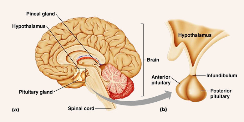
Hormones and the endocrine system
Usually work in parallel with the autonomic NS.
The endocrine system controls the secretion and production of hormones to be sent to certain parts of the body via the bloodstream.
An important one is the PITUITARY GLAND, which releases hormones that controls the production of other hormones wow. Its controlled by the hypothalamus in the brain.
The anterior pituitary gland is responsible for the production of ACTH, which is the main stress hormone.
Its responsible for stimulating the adrenal cortex to release corticosteroids such as CORTISOL.
Cortisol is released under long -term stress.
It is responsible for: lowering sensitivity to pain, glucose production?, lowering immnue response,
THEREFORE long-term stress can cause illness and possible coronary heart disease
NEURONSSSSS
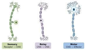
Between Receptors and the CNS
Between muscles and the CNS to allow movement yo
Between sensory and motor!
Synaptic Transmission
-
Synapse is a gap in the junction
- Neurotransmitters from the pre-synaptic neuron travel across the synapse to be received by the post-synaptic neuron
- The neurotransmitters bind to specific receptors on the surface of the post neuron.
- They are absorbed by the neuron or broken down by enzymes in the synapse.
- The nerve impulse is passed on this way along neurons until it reaches where its supposed to.
- Can be excitatory (increase likelihood of firing) or the opposite.
A2 JAZZ
SSPEC
Split brain research
Localisation of function in the brain and Lateralisation
The brain after trauma
Ways of studying the brain
Biological rhythms
Localisation of Function in the Brain
The idea that specific centres of the brain are responsible for specific functions.
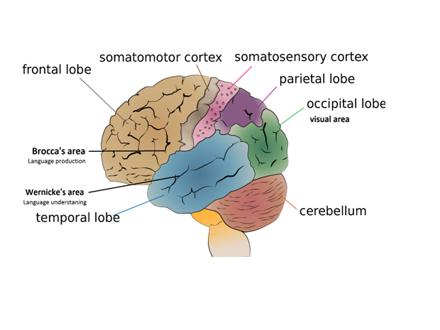
Motor Cortex
Control of muscles and movement in the body
It's in the frontal lobe
Contralateral control

Somatosensory Cortex
Detects sensory events happening in different parts of the body.
Can feel touch, pressure, temperature and pain and can localise where in the body this is occurring
Considered a part of the parietal lobe. duh
Function is contralateral.
Auditory Centre
Sound goes to the cochlea, in the inner ear
Nerve impulses travel via an auditory nerve to the brain stem
Decoding takes place in the brain stem: impulse? goes to the thalamus which decodes more and sends what it has? to the auditory cortex in the temporal lobe
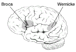
Broca's Area
Responsible for speech production and the muscle movements involved in speech production.
Located in the left hemisphere in the posterior section of the frontal lobe.
Damage to Broca's area results in difficulty producing speech and writing. (Production aphasia)
Paul Broca studied a patient nicknamed ‘Tan’ because, although he could understand language, ‘tan’ was the only word he could say or write. When examined, post-mortem, Tan had a lesion in the area of the brain now known as Broca’s area, and so did 8 other patients,
with similar speech deficits, studied by Broca.
Wernicke's Area
Responsible for processing and understanding speech.
Found in the temporal lobe.
Damage can usually lead to the inability to understand speech, while the ability to respond fluently still remains. (receptive aphasia)
Visual Centre
Light enters retina at the back of the eye
Nerve impulses are sent to the brain via an optic nerve
Goes to the thalamus (most stop here)
Then sent to the visual cortex in the occipital lobe.
The visual cortex spans over two hemispheres, as with most parts that do this, there is contralateral control.
The right peripheral vision is processed by the left side of the brain, and vice versa.

Evaluation of Brain Localisation
A lot of supporting evidence-
e.g the case of Phineas Gage, who suffered a motor accident where a pole went through his head, his personality changed.
this provides evidence that the part of his brain that was damaged must have been related to mood regulation.
This is supportive, however the method used to research PG was questionable, they asked his friends how he was before the accident, may not be the most accurate way to determine personality change
Evaluation of Brain Localisation
We have better, more scientific ways of knowing how the brain works
COME BACK
Evaluation of Brain Localisation
There is evidence for Wernicke's and Broca's areas from brain scans.
Peterson and et al found that Broca's area was active during reading tasks whereas Wernicke's area was active during a listening task. This demonstrates that these areas of the brain have different/seperate functions.
What's more, damage to these areas specifically leads to aphasia
~explain how that works~
this demonstrates the validity of assigning function to parts of the brain.
Evaluation of Brain Localisation
HOWEVER
Evidence from Lashley suggests otherwise. Her theory of equipotentiality suggests that different parts of the brain can take over the function for those that are damaged.
This view is an opposing limitation that is supported by research into functional recovery. It has been found that in the brain of stroke victims, the brain can re-wire itself over time and that patients can regain some function.
This goes against the theory of localisation, and shows that different areas of the brain may be able to perform several fucntions
Brain Lateralisation
Research has shown that the right and left hemispheres of the brain are responsible for different functions.
Right- Creativity, spatial ability, context, recognition of faces, places and objects
Left- Speech production and comprehension, analysis and calculations, time and recognition of words and stuff
Split Brain Research
Split brain patients have had their corpus collosums cut, usually due to epilepsy. This means there is no communication between the right and left hemisphere.
SPERRY set up an apparatus that would allow a stimulus to be sent to just one visual field at a time. Ppts were blindfolded on one eye and info was sent to either visual field in the other.
They looked at a screen, which would flash an image or word.
Participants were asked to respond either verbally, or with either hand. Both hands are covered.
Results and Conclusions?
Basically, images flashed to the right visual field go to the left hemisphere, because of lateral control.
The left hemisphere is in control of language, so images flashed to the left visual field (that go to the right hemisphere) would be unarticulatable
If an object is seen from the right visual field, the right arm would be unable to pick up the correct object.
If one word was flashed in each visual field, the participant would say the thing in the right visual field and write the one in the left.
This research shows that the two hemispheres are responsible for different things. it doesnt show localisation of function, just shows a connection between the two hemispehres is as important as each part
Split Brain Research EVALUATION
SPERRY used only 11 participants
- Some had experienced drug therapy longer than others, so it's unclear whether his results were skewed by variations within the group
-On the other hand it allowed him to gather rich quantitative and qualitative data since the sample was so small.
Ecological validity
- Apparatus and method were highly reliable, participants blindfolded etc.
- This is an issue because in real life, split brain patients use both eyes. the other eye usually compensates for the lack of corpus collosum
Split Brain Research EVALUATION
Lateralisation appears to change throughout or lifetime
-Research has shown that lateralisation of language becomes pronounced in children and adolescenets but decreases every decade after the age of 25.
This could be because brain function deteriorates with age, so when you're older, different parts from each hemisphere have to compensate function.
this means its oversimplifying to say that brain lateralisation is permanent
Split Brain Research EVALUATION
Case studies challenging
- JW developed the capacity to speak out of the RIGHT HEMISPHERE!
therefore it is incorrect to assume that language control is only in the left hemisphere.
THE BRAIN AND TRAUMA
Plasticity and functional recovery
What is Trauma?
Traumatic brain injury occurs when an external mechanical force causes brain dysfunction. Traumatic brain injury usually results from a violent blow or jolt to the head or body.
Can result in cognitive, behavioural and psychological impairment
Plasticity
The brains ability to change and adapt based on our experiences and new learning
Resarch into Plasticity
GOPNICK ET AL- Found that the number of synaptic connections in the brain peaks at 15,000 around 2-3 YEARS OLD.
Twice as many as the average adult brain.
the decline is due to SYNAPTIC PRUNING , in which rarely used connections are deleted.
MAGUIRE + WOOLETT (The Knowledge)
Used MRI scans on 16 Black Cab drivers and 50 controls.
The cab drivers had all passed the Knowledge, which is an extensive memory test of thousands of roads and POIs around London.
They found increased grey matter in the POSTERIOR HIPPOCAMPI in both hemispheres than the control group.
A positive correlation was seen between increase of size and time spent as a taxi driver.
This suggests that extensive time with spatial navigation can affect the hippocampus PHYSICALLY.
DRAGANSKI
MRI SCANS at 3 different points of a medical students life lol.
3 months before, day after and 3 months after medical exam.
During the first three months, the grey matter increased significantly in the parietal cortex bilaterally
Changes remained after 3 months.
* Increased grey matter was more prononuced 3 months after than the day after, so volume continued to increase.
Learning a large amount of information can LEAD TO STRUCTURAL CHANGES IN THE BRAIN.

FUNCTIONAL RECOVERY
A form of plasticity, in which different parts of the brain will adopt the function of another if that part was to get damaged.
Research into Functional Recovery
The two ways in which the brain can recover functionality are neural unmasking and stem cells
The brain creates new synaptic connections in order to compensate for the damaged area of the brain.
Neural pathways are 'unmasked' to enable function to continue:
Axonal Sprouting: new nerve endings grow and connect with undamaged neurons to make new pathways
Reformation of blood vessels
Recruitment of homologous areas: Corresponding areas of the brain on the other hemisphere would carry out the same function.
NEURAL UNMASKING
The unspecialised cells can be injected into the brain so as to adopt the role of damaged neurons/ brain cells.
They can also secrete growth factors that can help rescue injured or damaged cells.
STEM CELLS
Applications in neurohabilitation
- Research shows that the brains ability to repair itself slows after a few weeks.
- We know now that further intervention is necessary: physio or electrical stimulation, to stimulate the growth of cells.
*important economic implications because it means that more elaborate, expensive solutions do not have to be considered.
Evaluation
There is evidence that plasticity occurs all throughout life, not just childhood.
Bezzola et al used ppts aged 40-60, found that there was reduced motor cortex activity during their swing after golf lessons compared to a novice control group. Shows that they learned, brains became more efficient - ADAPTED from an external stimulus. Also supports the use of fMRI scans..
Evaluation
BUT there are individual differences in the brains ability for functional recovery.
SCHNEIDER found that the more time an individual spent in education, the more likely they were to have a DFR (disability-free recovery) after brain trauma.
People with university-level education were 7x more likely to achieve DFR.
This is evidence for a 'cognitive reserve', which occurs as a result of education.
However, DATA COLLECTION was retrospective and no IV was manipulated, so we cannot know if results are directly/only linked to education.
Support from animals studies
TAJIRI injected rats with brain trauma with stem cells.
Three months later they showed recovery and clear development of neuron-like cells. The same was not seen in the control group. This shows the effect of stem cells and demonstrates their usefulness.
However, stem cells are a controversial area of treatment, not easily come by, animals.
Evaluation
Ways of Studying the Brain
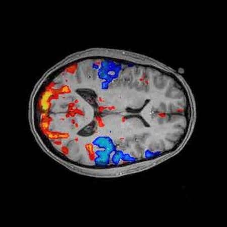
fMRI Scans
How does it work?
fMRI machines detect changes in blood oxygenation and blood flow that occurs in the brain as a result of neural activity.
When areas of the brain are active, they require more bloodflow for oxygen. This is known as the haemodynamic response
What Does it show?
A 3D image showing which part of the brain is active during activit
For whom can you use it?
-
For soft tissue (the brain for example)
-
Bodies with no metal in them
-
People who do not have claustrophobia
-
If you wanna study internal mental processes
Advantages
- Provides a 3D image of bloodflow
- No radiation (unlike PET)
- Non-invasive
- Risk free
- High spatial image
Disadvantages
- Expensive
- Poor temporal resolution (5-second lag) between image on screen and firing of neurons
- Only shows bloodflow, not actual neural activity or individual neurons
Electroencephalograms
EEGs
How Does it Work?
Electrodes are placed on the scalp.
Electrical activity in the brain is measured for a certain length of time.
Measures the activity of many electrons firing at once, not individual ones.
What does it show?
Shows overall electrical activity.
Patterns of waves are produced that represent levels of arousal or consciousness at different times and in response to different stimuli.
What is it used for?
-
Used in sleep studies to measure the stages of sleep
-
As a diagnostic tool: abnormal EEGs can be seen in patients with epilepsy, anorexia, depression and schizo.
Advant
- Valuable for detecting epilepsy
- Contributes to our understanding of sleep
- Has really good temporal resolution
- Non-invasive
- Cheaper than fMRI
Disadvant
- Doesnt show individual neurons firing
- Poor spatial resolution, unable to tell where the brain waves are coming from.
Event-Related Potentials
How does it work?
Using EEG's means ending up with a vague and general show of brain activity.
Within all the data however are waves that are specific to a certain stimuli.
Using a statistical averaging technique, researchers can ilter out the waves made from unwanted stimuli
What remains are the even-related potentials specific to a certain stimulus
Advantage?
They make it possible to see someone's response to something without them having to actually respond.
If the patient cannot respond due to inury
But
Not all voltage channes across the scalp are recordable.
Electrical activity from deep in the brain is not recorded. This means that, at current, ERPs are restricted to the neocortex, the most recently evolved part of the brain
Post-Mortem Examinations
How is it done?
Involves removing the brain from the skull (after death) and dissecting it to look at abnormalities when compared to a neurotypical brain
It's usually done on people with rare disorders to see how their brains differ from a neurotypical one
Good
Allow for a more detailed examination of the brain than non-invasive techniques. Researchers can look at deeper parts of the brain such as the hypocampus and hypothalamus
THis is useful in cases such as that of HM, who's post-mortem showed his memory problems were due to lesions in the hippocampus
But
There can be lots of confounding influences on the brain,
cause of death, age, drug use, length o time between death and exam. these could all influence the anatomy of the brain and prevents us from establishing cause and effect between abnormalities in the brain and abnormal behaviour
BIOLOGICAL RHYTHMS
ULTRADIAN RHYTHMS
Less than 24 hours
Eating, rest-activity cycle, stages of sleep
Research into Ultradian Rhythm
McClintock and Stern
10 year long longitudinal study
29 women who had irregular periods. 9 of them had their sweat swabbed and put on the upper lip of the other 20 participants
68% of the time the participants who had recieved sweat found their cycles closer to that of the donors.
Infradian rhythms can be affected by external factors as well as internal ones.
Research into Ultradian Rhythm
Seasonal Affective Disorder
SAD is a depressive disorder which has a seasonal pattern of onset – throughout the winter months. Its symptoms are general low mood and lack of interest in activity. At night, low light levels stimulate the production of melatonin, this is what triggers sleepiness. Therefore, you would expect the lower light levels of the winter months to have a similar effect. In areas where light levels are exceptionally low for prolonged periods, such as the Polar regions, you would expect the effects to be particularly noticeable. Terman (1988) found that SAD was five times more common in New Hampshire, a northern state of the USA, than in Florida, obviously a sunnier clime. SAD could also be considered a disruption of a circadian rhythm since it could be seen as due to disruption of the sleep/wake cycle during the darker winter months.
CIRCADIAN RHYTHMS
Last around 24 hours
Sleep wake cycle, core body temperature, hormone production (melatonin)
Circadian rythms are synchronised by an internal pacemaker, the suprachiasmatic nucleus (hypothalamus) (or SCN)
The pacemaker needs to be constantly reset so that it is in sync with the outside world. Light acts to provide that reset.
Our 24 hour sleep cycle is influenced by patterns of daylight and darkness. These are exogenous zeitgebers.
These help to maintain the rhythm but are not necessary. Our circadian rythm is 'free-running' and does not require exogenouses in ourder to keeo a 24-25 hour cycle
Research into Circadian Rythms
Siffre was a cave spealist who lived in a cave for ~6 months so as to investigate the effect of having no exogenous zeitgebers.
He lived in a pitch dark cave with only a lamp, food water, a means of communicating with the outside world, but not clock or watch.
He slept and ate whenever he felt like it.
He found that his sleep-wake cycle settled on 25 hours naturally, showing that without light, his body ran its natural clock
Research into Circadian Rythms
Folkard
Had 12 participants stay in a cave for 3 weeks with no way of telling the time besides a large clock controlled by the experimenters.
The participants agreed to go to bed at 11 45pm and get up at 8 45pm.
At first the clock worked at a 24 hour pace and then they sped it up to a 22 hour pace
Only one of the participants adapted their cycle to the clock, the rest kept their 24 hour sleep-wake cycle.
so dont overestimate the role of the EXTERNAL crap, endogenous pacemakers are important too
Evaluation of Circadian Rhythm research
- Practical applications
- Research has shown that the risk of heart attack is the most in the morning so drugs have been developed that can be taken at night to be released in the morning
- And, shift workers have been seen to have more accidents around 6am when their concentration reduces. Night shift workers are also 3 times as likely to develop heart disease due to the stress of trying to adjust to different sleep/wake cycle.
- Problem is that the sample sizes used for these studies is usually quite small, so cant apply results to general population or anything. Siffre himself said that his sleep/wake cycle slowed down as he aged, which shows that there can be individual differences between participants.
Evaluation of Circadian Rhythm research
Lack of control of extraneous variables
Siffre had a small source of artificial light with him, which could have acted as an exogenous zeitgeber. Czeisler found that even artifical light can affect circadian rhythms. So we cannoot e sure therefore if the circadian rhythm is free-runnning and 24/25 hours long
INFRADIAN RYTHMS
Last longer than 24 hours
Menstrual cycle, animal migration and hibernation
Endogenous Pacemakers
Superchiasmatic nucleus: found in the hypothalamus. Controls and syncs all other clocks in the body
Pineal gland: releases melatonin throughout the night to make us sleep (in low levels of light). And decreases it during the day
Exogenous Zeitgebers
Light: light stimulates the light receptor cells in the eyes. Breaks down a melanopsin, which sends impulses to the SCN.
Social Cues: things such as meal times and social activities can act as exogenous zeitgebers that tell the body when to be awake and when to sleep.
BIOPSCHOLOGY
By Zubiya Burney


