Student Experience
STFC/Durham University CDT in Data Intensive Science.
Carolina Cuesta-Lázaro
Arnau Quera-Bofarull
(Joseph Bullock)
Placement at IBEX Innovations Ltd.
Who are we?


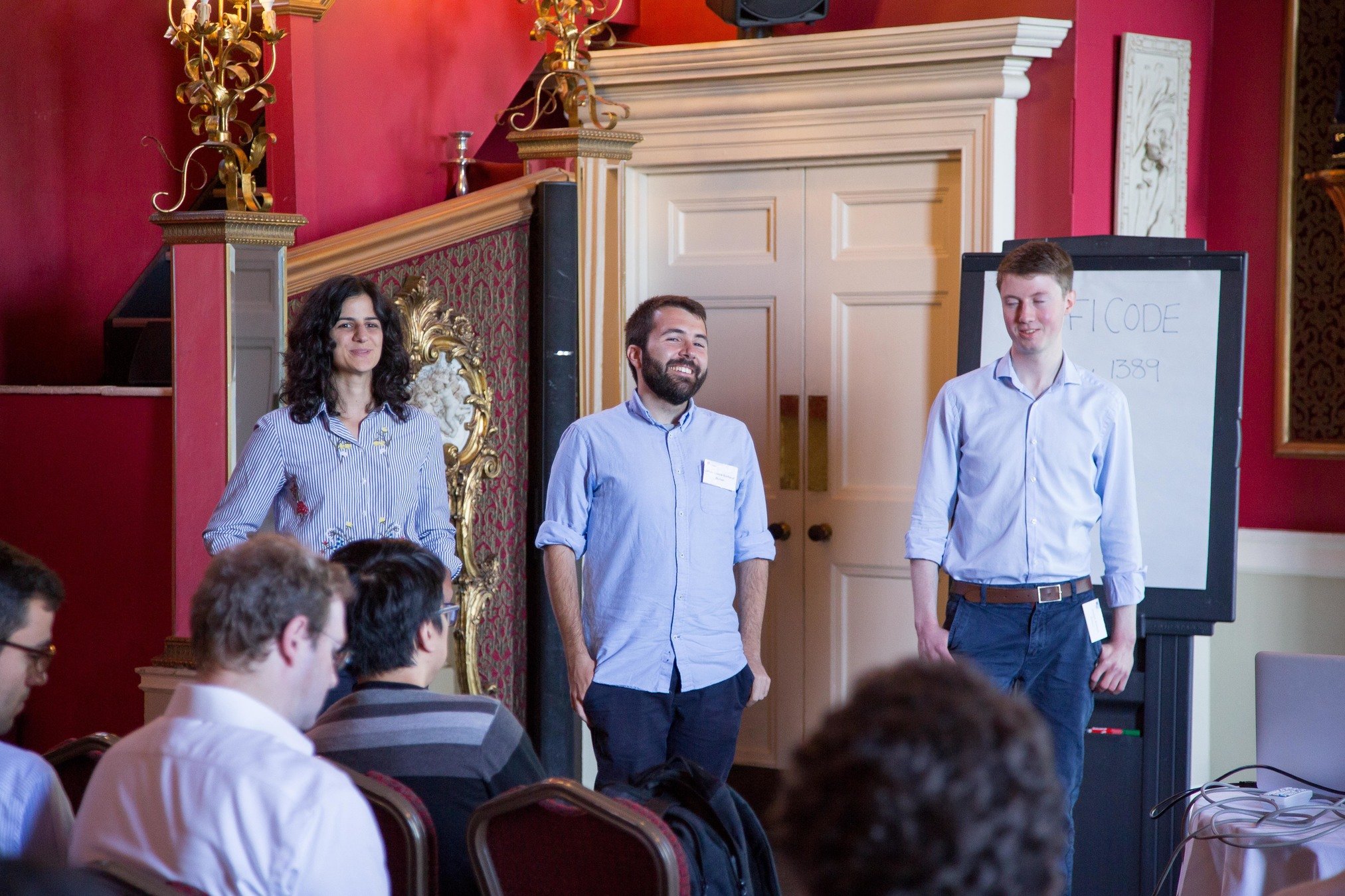
2 months team project at IBEX innovations
Carolina / Arnau
Cosmology
Joe
Particle Physics
Detect bone and soft-tissue on X-Ray images
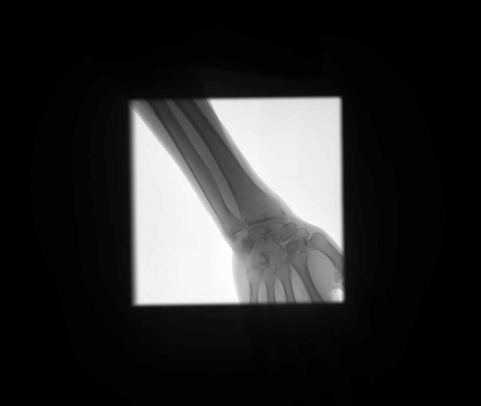
Detect
collimator

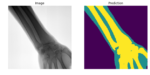
Segment

Open beam

Bone

Soft-tissue
Previous approaches
- Challenging problem due to varying brightness throughout the image.
- Usually done by detecting edges and shapes.
- High accuracy requires tuning of hyper parameters per image and body-part (not automated).
- Not well defined boundaries.
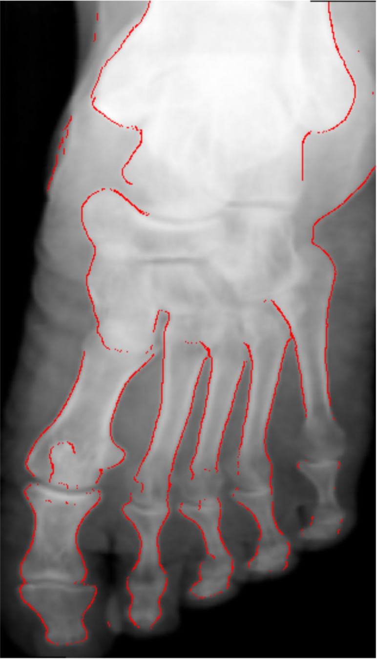
Kazeminia, S., Karimi, et al (2015)
Are there better features hidden in the data?
Extracts features from a high dimensional feature space, once trained on a particular dataset.
DEEP LEARNING

What is training?

Luminosity
Size
Colour
Galaxy
Star
x 2

LOSS = GENERATED OUTPUT - ACTUAL OUTPUT
Galaxy
Star
x 4
Deep learning on images
Credit : https://www.pnas.org/content/116/4/1074

With the right data, the network will find the right features
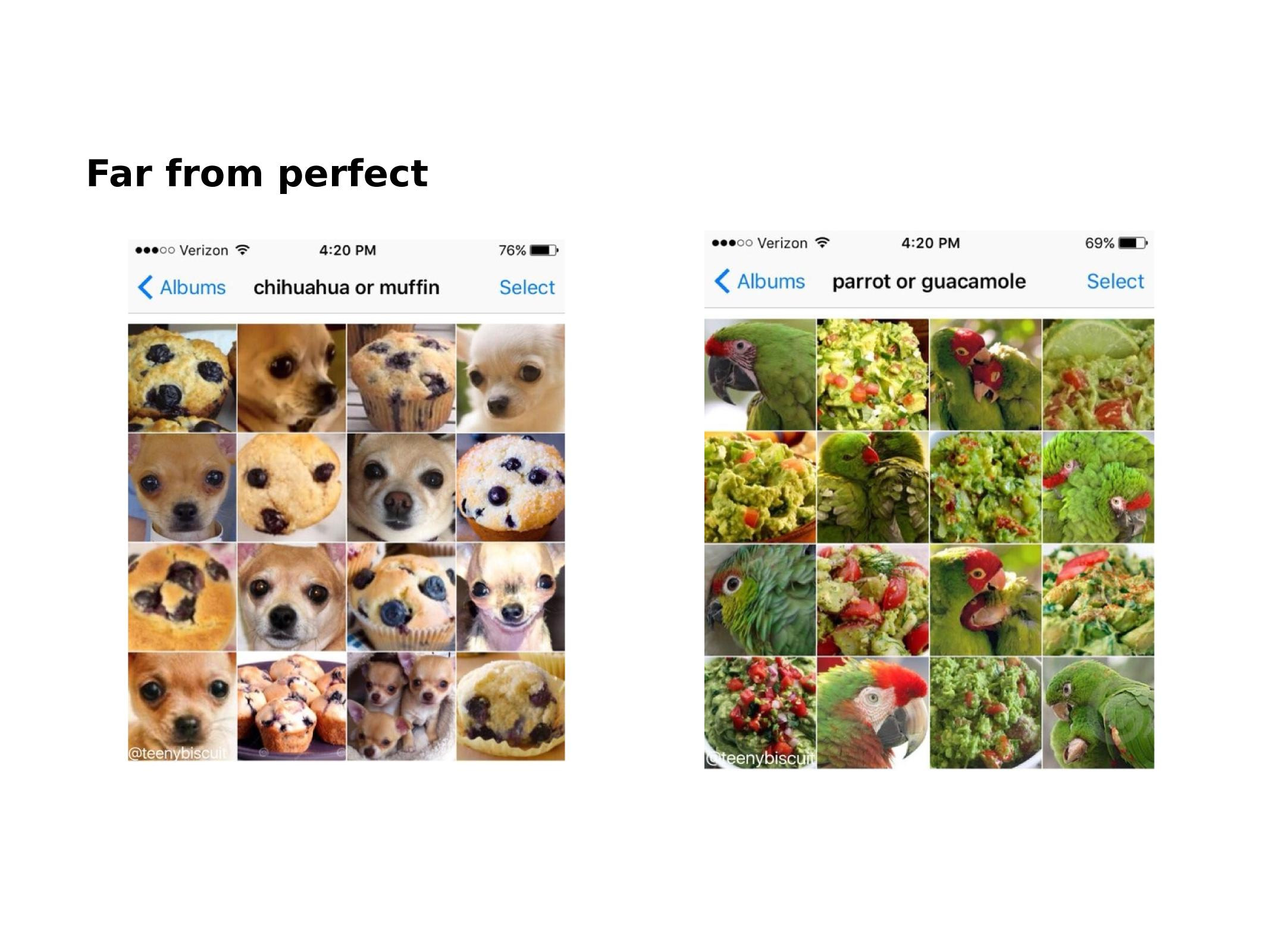
With wrong data ....
Does it really work?
SegNet
- The network has more than 15 Million free parameters.
- To find the values of the parameters that produce the correct segmentation, it has been trained on 1.3 Million images.

Could it solve our problem?
150 labeled images.
Hardware limitations (memory, training time...).
We need a fast network, easier to re-train as we get more images.
CONS
PROS
Could work for different detectors (different noise).
Generalize to different body-parts.
Well defined boundaries between regions.
Could be improved through more training.
The Road to XNet

Coursera
The Dataset
- Small, ~150 images
- Unbalanced
Solution:
Artificially augment the dataset by transforming original images.

Splitting the dataset
We typically divide our dataset into three subsets:
- Training: 70% from categories with more than one sample.
Data augmentation -> Equal sample size for all categories.
Final size ~ 7000 images. - Validation: 15% from categories with more than one sample. Used to stop training and hyperparameter tuning.
- Test: 15% including categories with only one sample. Final network performance is evaluated in this set.
Network Architecture
First attempts focused on a very simplified SegNet model.
Underfitting
Network Architecture
Going deeper has limits ( limited image size, GPU memory bottleneck, overfitting).
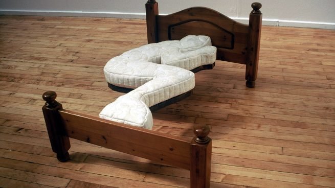
Overfitting
Dealing with overfitting
Ways to reduce overfitting
- Increase the dataset.
- Reduce network complexity.
- Regularisation.
Idea:
Penalise the network if it uses too many parameters to fit the data.

Credit: www.kdnuggets.com
XNet
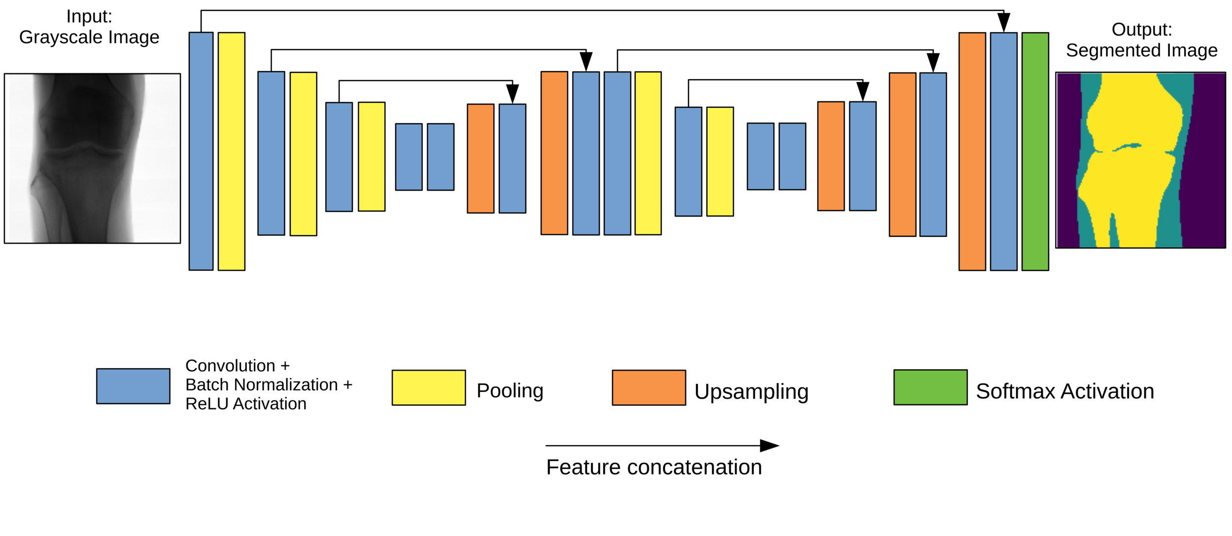
- Typical encoder-decoder architecture.
- W-shape for two feature extraction stages. Avoids resolution problems.
- Skip connections across levels.
- L2 regularisation at each convolutional layer.
Final model: XNet
Post-processing
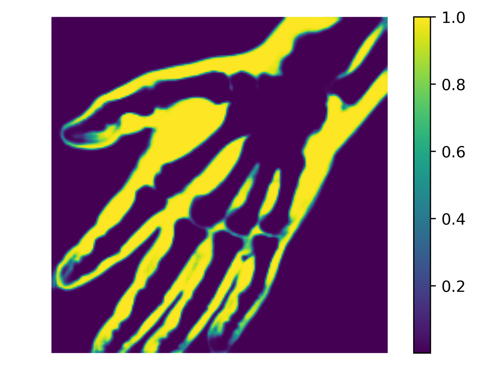
The network outputs 3 probability maps.
Soft tissue probability map

We can reduce the number of false positives by making a probability cut to the map.


Probability
Results

- Generalises well even for unseen categories!
- Overall accuracy on test set: 92%
- Soft tissue TP/FP rate: 82% / 4%
Comparison with other methods
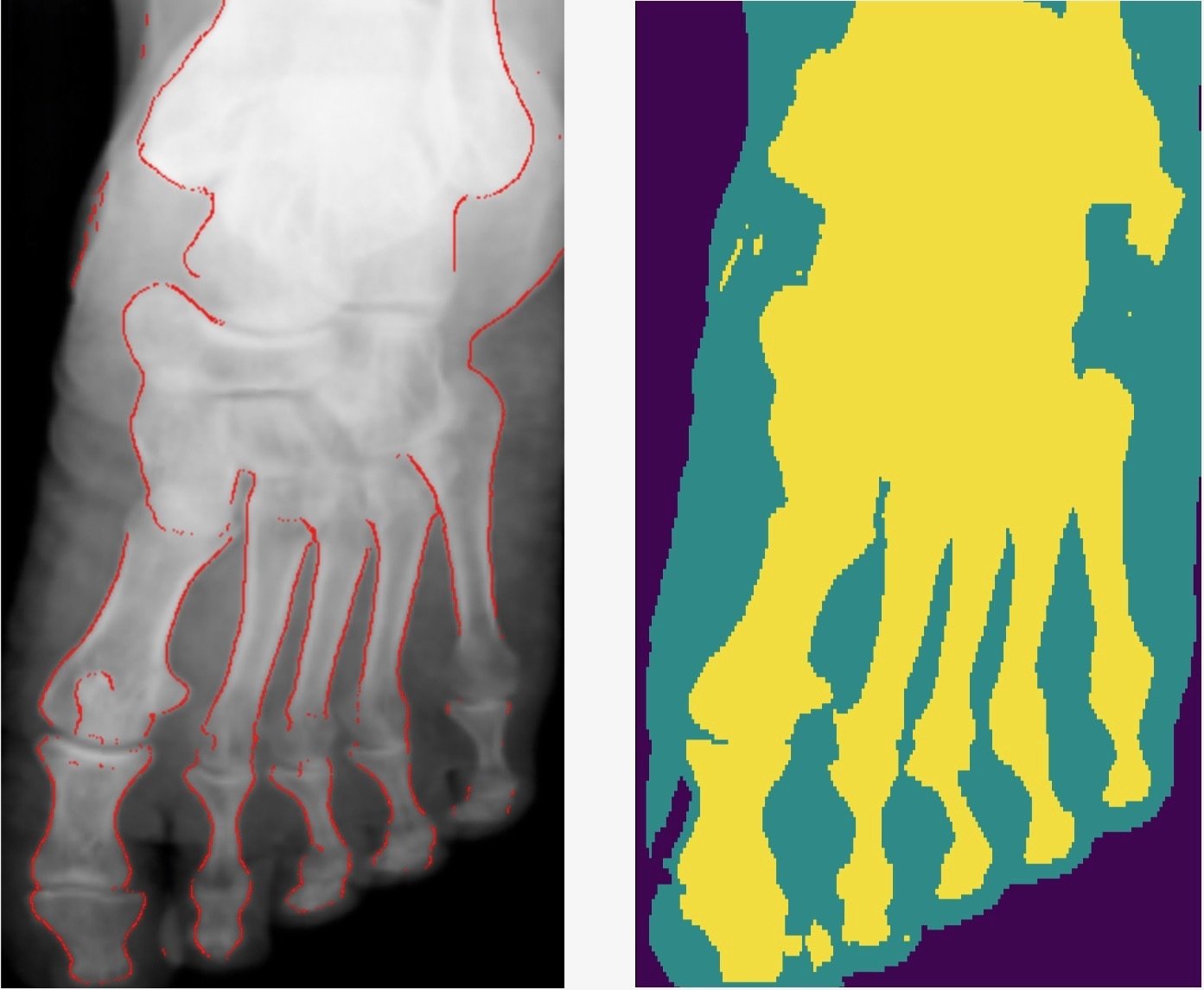
- Smooth connected boundaries.
- Better generalisation to different body parts (we do not have any frontal view of a foot in our dataset).
- More robust to noise.
- Well defined metrics to benchmark against.
- The development process takes a long time due to hyperparameter tunning (50% of our internship time).
-
We used ~1000 GPU hours.
- 3x 4GB GPUS
- 1x 8GB GPU
- AWS 12GB GPU
Development process

Cryptocurrency Times
- Promising ML applications to medical imaging.
- Possible to train ML models with limited hardware and resources.
- Knowledge of building and deploying a machine learning product in an industrial setting.
-
Paper out arXiv:1812.00548v1, and presented at the SPIE Medical Imaging conference in San Diego. Best student paper awarded.
Conclusions
Learning outcomes
beyond-the-lab
By arnauqb
beyond-the-lab
- 994


