Lab 10

Agenda

  1. Turn in Lab 9 by the staplers on the North bench.
    1. Needs a name and group number
  2. ~10 min.: Q&A on Lab 9 material
  3. 20 min.: Quiz 9
  4. ~2 hrs.: Lab 10: ECG and Blood Pressure
  5. ~30 min.: Next lab reminders/questions/review

Lab 9 Q&A

Blood Components

Normal Blood Values

  • Hematocrit
    • ♂ 45%-52%
    • ♀ 37%-48%
  • Erythrocyte count: 4.5-5.5 million/microliter
  • Hgb: 14-16 grams/100 mLs
  • Leukocyte: 5k-9k/microliter
  • Platelets: 200k-500k/microliter

Blood Types

Erythroblastosis

Quiz 9

ECG and Blood Pressure

Lab 10

Lab Purpose

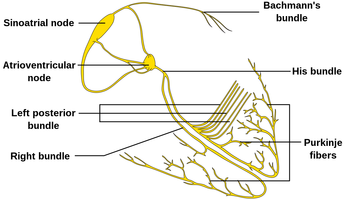
To allow students to experiment with principles of blood pressure regulation, cardiac cycle, and the interpretation of electrocardiograms (ECG).

Lab objectives

  1. Name all of the components of the normal ECG and describe the electrical event that corresponds to each deflection or interval relative to the (+) ECG electrode.
  2. Describe the pathway of the action potential through the heart from beginning to end.
  3. State the function of the SA node and AV node in electrical conduction through the heart.
  4. Understand the mechanism of auto-rhythmicity in cardiac muscle tissue
  5. Measure the ECG, PR interval, Q-T interval, QRS width, etc.
  6. Describe ECG changes that occur with increased heart rate with each component of the ECG.
  7. Describe the events corresponding to the heart sounds and how they are correlated to the QRS complex.
  8. Measure blood pressure and calculate pulse pressure and mean arterial pressure.

Lab Safety

How to use the ECG or EKG

Measurements on EKG

Small squares are 0.04 sec. wide

Large squares are 0.2 sec. wide

How to take blood pressure

Procedure Hacks

  • Ladies - if you're getting the ECG, you should go to the bathroom to put the electrode patches on.
  • Print out one copy of the ECG. Mark it up and then give it to me to make copies.
  • Do not tear away EKG leads. Don't lose parts!
  • When getting EKG data, get it as fast as possible (i.e. right after the subject stands or gets off the bike).

Core Principles

Name the components of an EKG wave

Name the components of an EKG wave

  • P wave = atrial depolarization that precedes atrial systole.
  • QRS complex = ventricular depolarization
    • (atrial repolarization occurs during this time, but is obliterated by the greater QRS complex)
  • T wave = ventricular repolarization that occurs just before the end of ventricular systole.
  • P-R interval = time between the beginning of the P wave and the beginning of the QRS complex.
    • Typically about 0.15 seconds, this reflects atrial depolarization (the P wave) and the time delay caused by the AV node for the electrical impulse to travel from the atria to the ventricles (represented by the flat area of the P-R interval.
  • Q-T interval = time from the beginning of the Q wave to the end of the T wave.
    • This normally lasts less than 0.38 seconds and approximately corresponds to the time of ventricular contraction.

How does an action potential travel through the heart?

  1. SA node - generates spontaneous AP
  2. Internodal tracts
  3. AV node - delays AP to allow for ventricular filling.
  4. Bundle of His
  5. Right bundle and left posterior bundle
  6. Purkinje Fibers

How does an EKG change with increased HR?

Pulse Pressure

Pulse \ Pressure = Systolic \ Pressure - Diastolic \ Pressure

Mean Arterial Pressure

Mean \ Arterial \ Pressure \, (MAP) = Diastolic \ Pressure + \frac{1}{3} \ Pulse \ Pressure

Cardiac Cycle

Lab 11: Respiratory Physiology

  • Read:
    • Silverthorn pages 535-559, 565-586
    • Stanfield pages 489-499
    Bring:
    • Calculator

Lab 10

By Ben Carter

Lab 10

Z00L-2425 ECG and Blood Pressure

  • 472