


Jesse Bloom PRO
Scientist studying evolution of proteins and viruses.
Fred Hutch Cancer Research Center / HHMI
Slides at http://slides.com/jbloom/mstp-2021
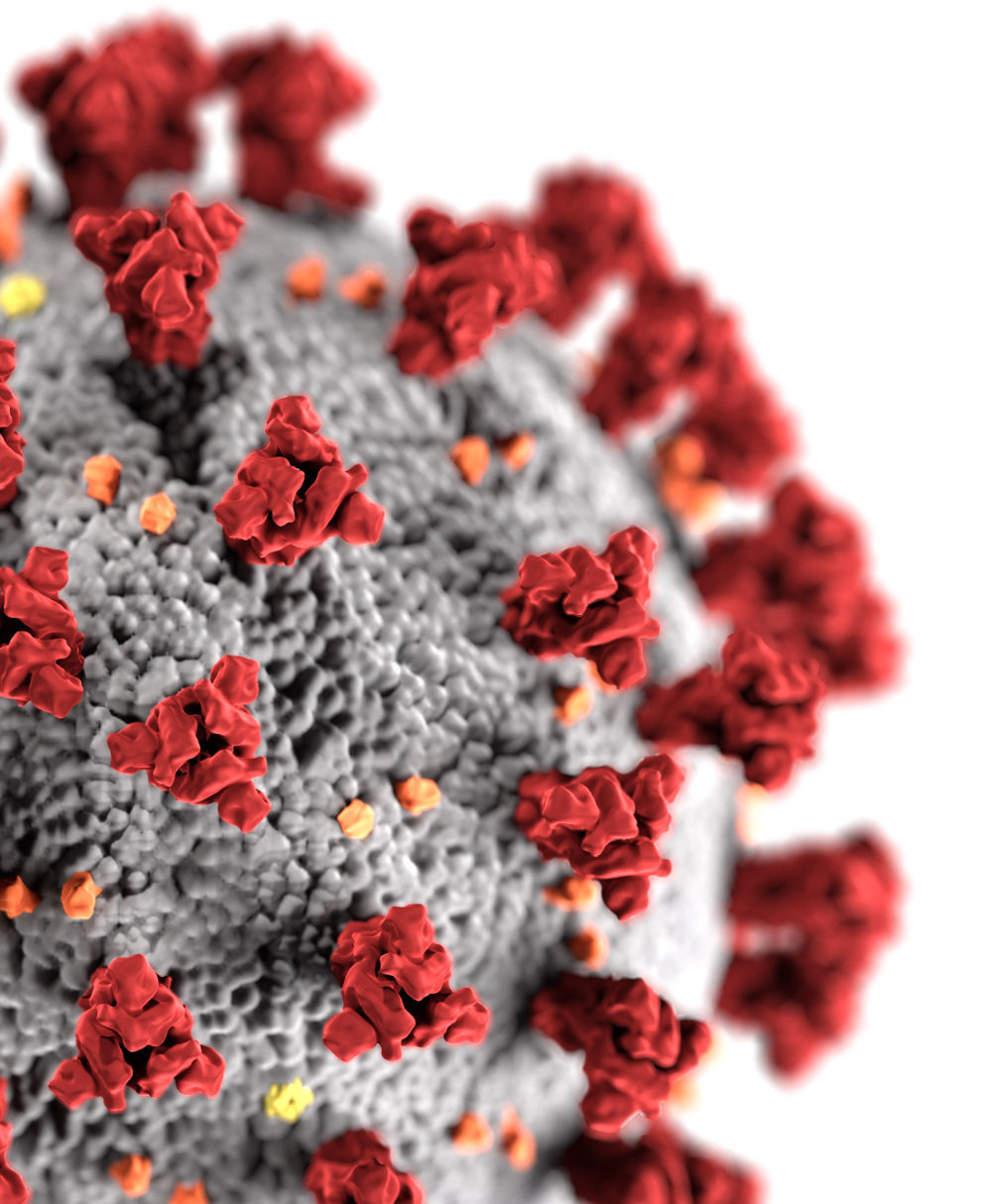
virion image from https://phil.cdc.gov/Details.aspx?pid=23312
-Asn-Ile-Thr-Asn-Leu-
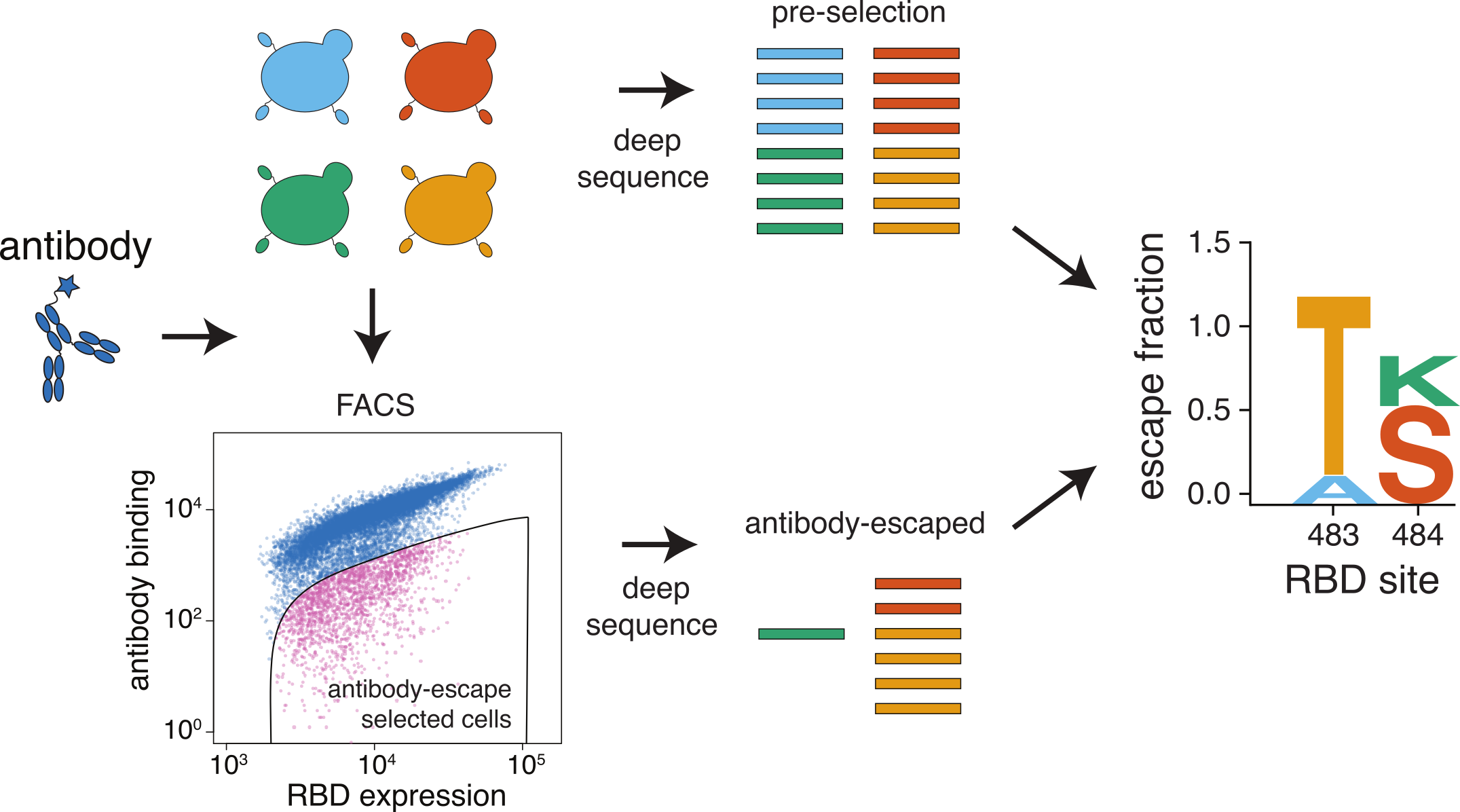
RBD phenotypes
- protein folding
- affinity for ACE2
- binding by antibodies
Structure still matters implicitly, as it largely determines mapping from mutation to phenotype.
-Asn-Ile-Thr-Asn-Leu-
-Asn-Ile-Thr-Glu-Leu-
-Asn-Lys-Thr-Asn-Leu-
(201 sites) X (19 amino-acid mutations per site) = 3,819 mutations
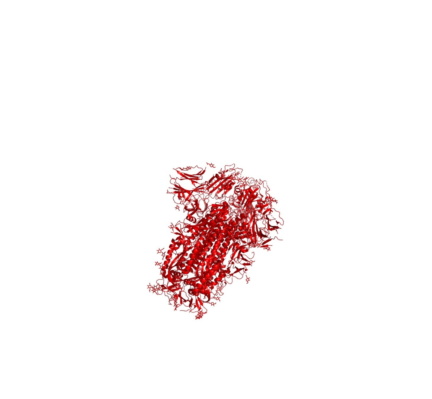
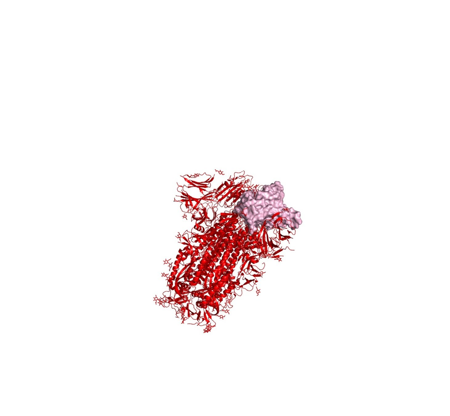
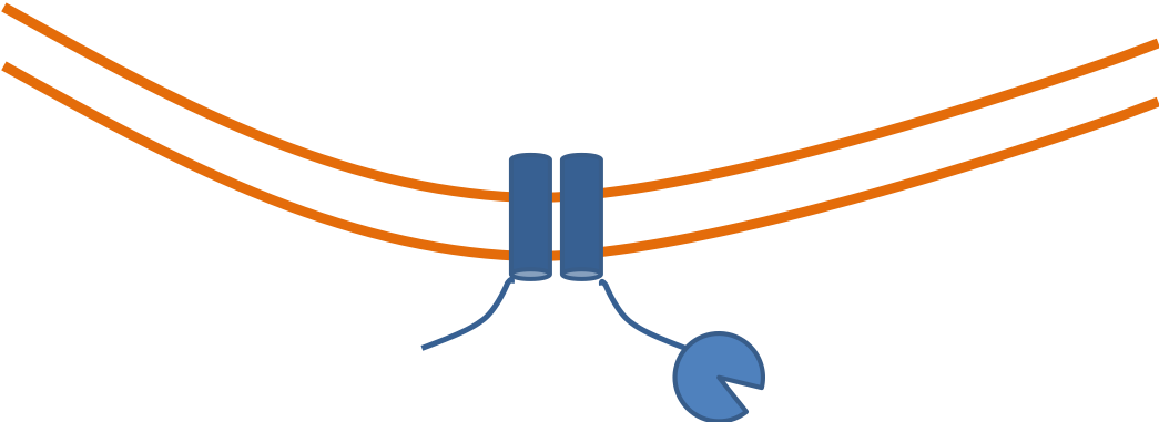
RBD
fluorescent ACE2
yeast
fluorescent tag on RBD
Click here for details on how library is made.
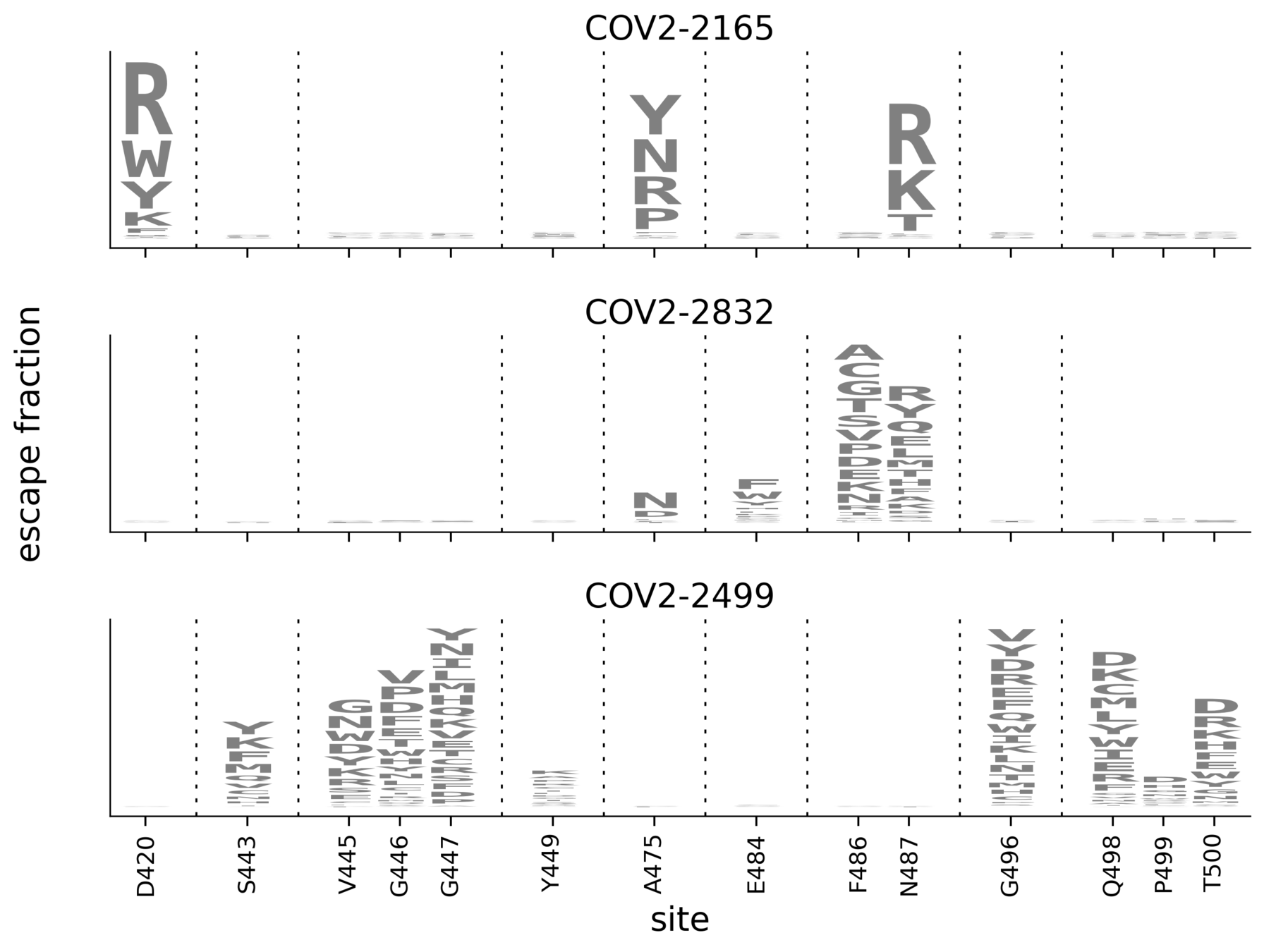
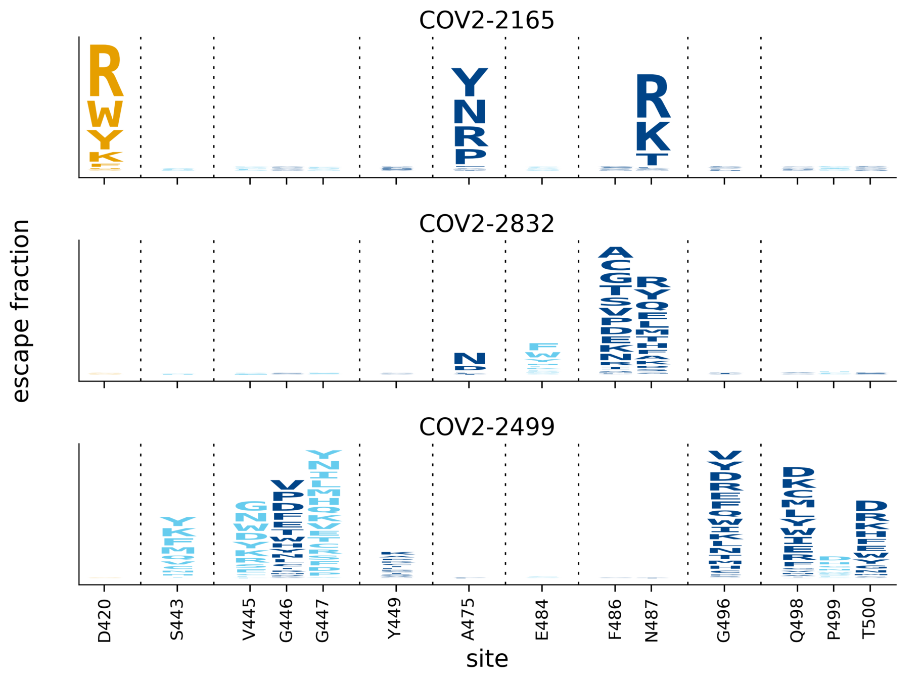
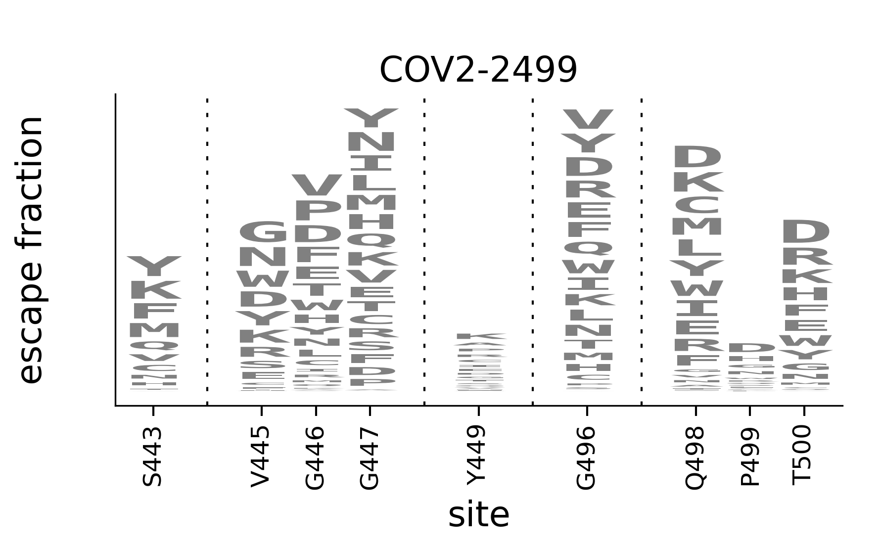
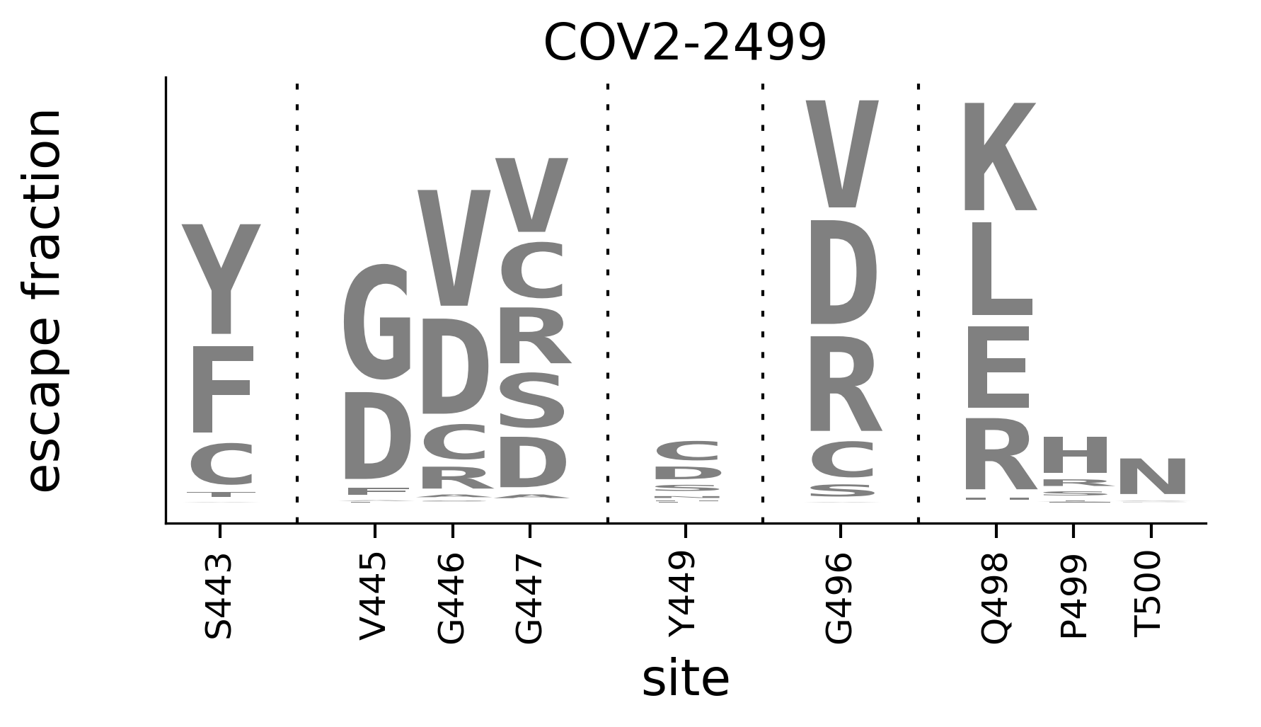
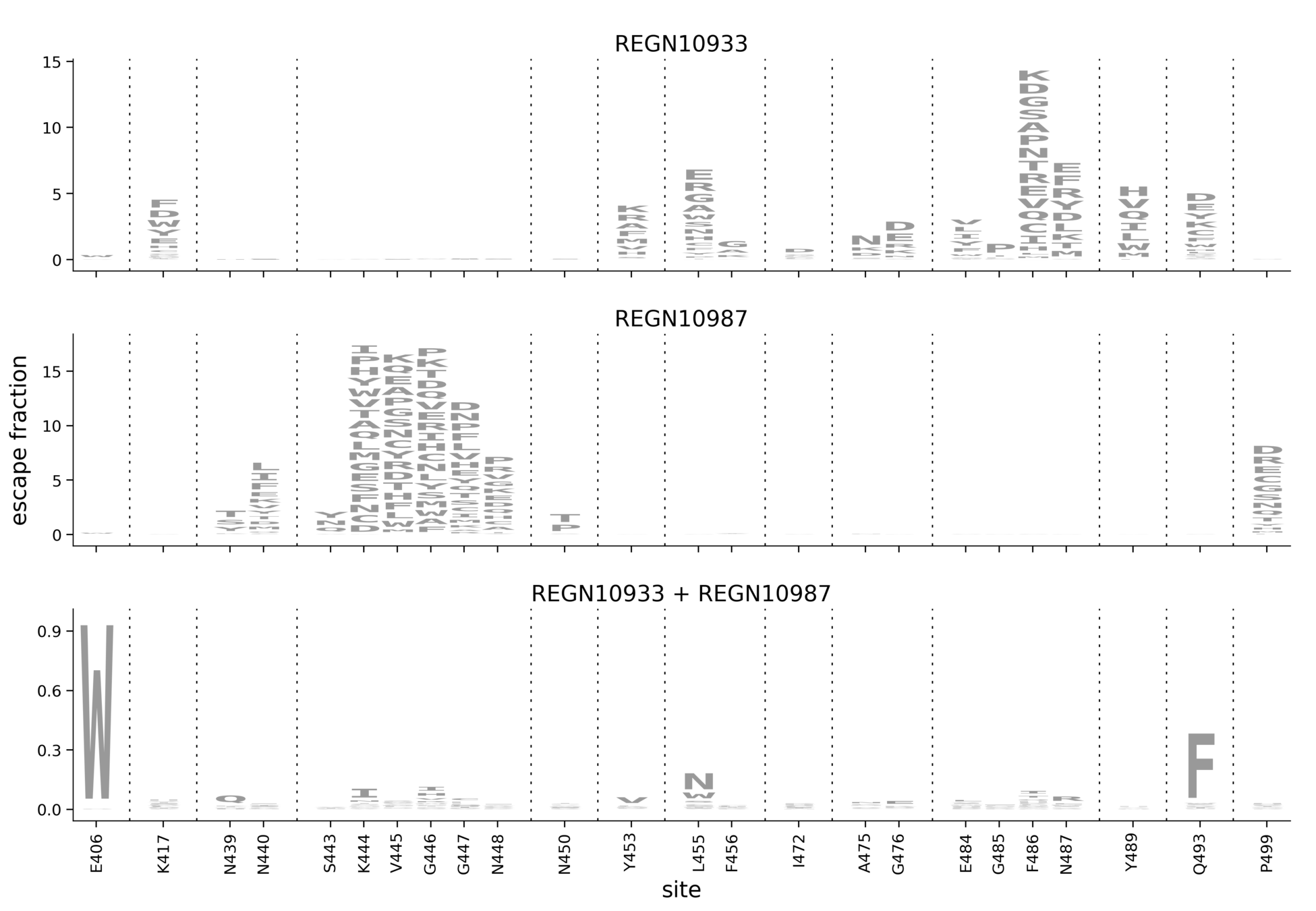
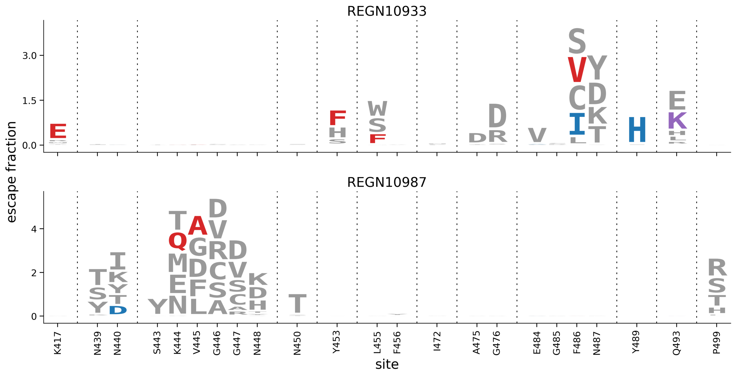
In maps, tall letters indicate strong escape mutations
core RBD
receptor-binding motif
ACE2-contact residue
core RBD
receptor-binding motif
ACE2-contact residue
Subtle differences between COV2-2165 & COV2-2832 (e.g., sites 486, 487) validate in neutralization assays (see here).
Spike-expressing VSV (Case*, Rothlauf*, ..., Whelan. Cell Host & Microbe, 2020) in a real-time cell analysis assay (Gilchuk, ..., Crowe. Immunity, 2020).
See here for more details.
mutation
G446D
Q498R
count
3
2
mutation
G446D
Q498R
count
3
2
mutation
G446D
Q498R
count
3
2
mutation
G446D
Q498R
count
3
2
ACE2 binding
weak strong
effect on escape
ACE2 affinity
effect on escape
ACE2 affinity
0 escape mutants in 56 attempts
RBD expression
ACE2 binding
RBD expression /
ACE2 binding
weak strong
Red are mutations from Baum, ..., Kyratsous. Science (2020) among single-nucleotide accessible mutations only
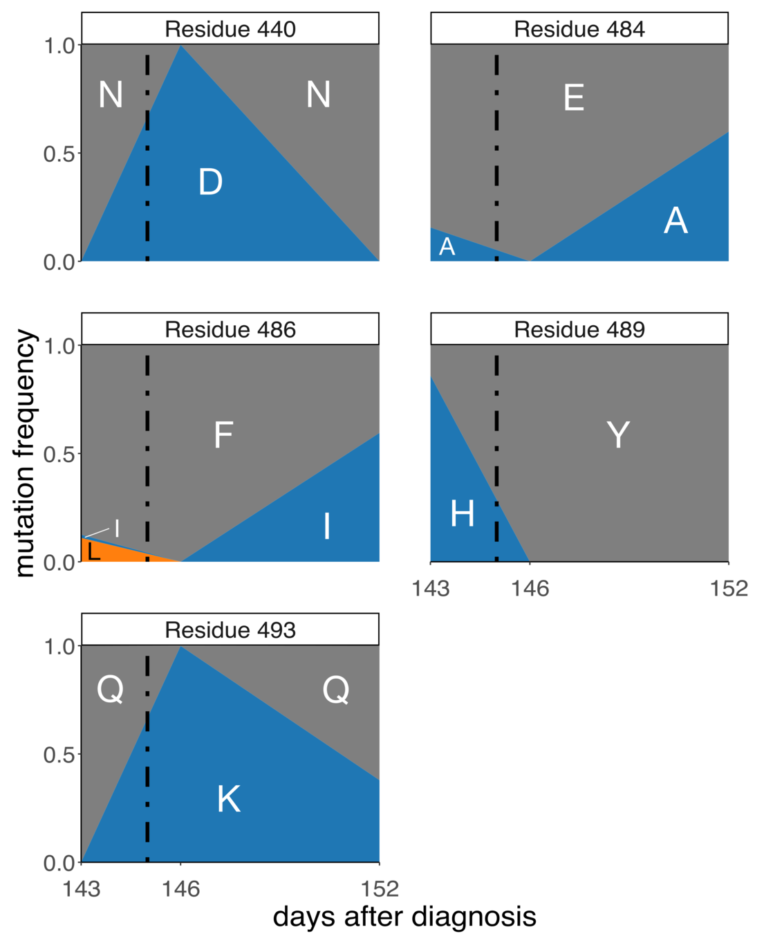
Collaboration with Jonathan Li, patient originally described in Choi et al, New England Journal of Medicine (2020)
Note extensive genetic hitchhiking and competition among viral lineages; also described in within-patient evolution of influenza in persistent infections: Xue et al, eLife (2017)
Regeneron cell-culture
Patient
Both
Most escape in patient not found in cell-culture selections, illustrates utility of complete escape maps
Tyler Starr
Allie Greaney
Sarah Hilton
Bloom lab (Fred Hutch)
Tyler Starr
Allie Greaney
Sarah Hilton
Bloom lab (Fred Hutch)
Li lab (Brigham & Women's)
Crowe lab (Vanderbilt)
Whelan lab (Wash U)
King lab (Univ Wash)
Veesler lab (Univ Wash)
These slides:
Interested in using the phenotypic maps? Please check out:
Other members of Bloom lab:
Kate Crawford
Adam Dingens
Will Hannon
Amin Addetia
Andrea Loes
Rachel Eguia
By Jesse Bloom
The evolutionary potential of the SARS-CoV-2 receptor binding domain


