題
cf 1045B 708C 1085F 1187F
min25 篩
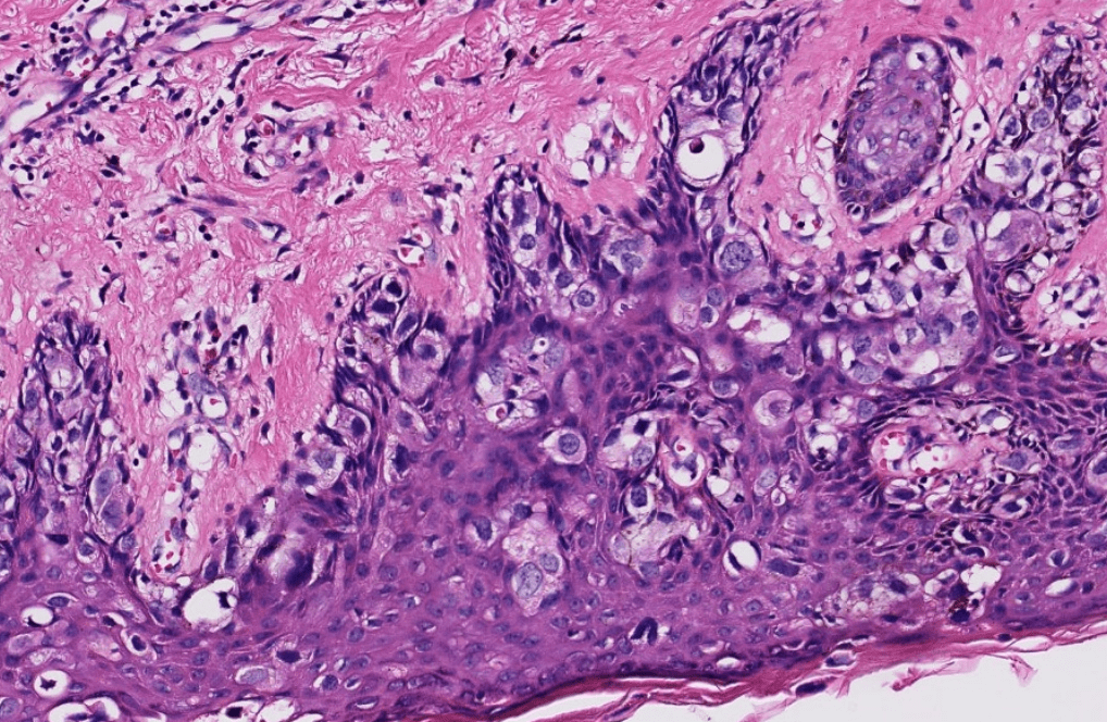
Pathology-Micro 4
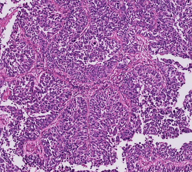
urothelium
may have squamous differentiation
loss of basal cells
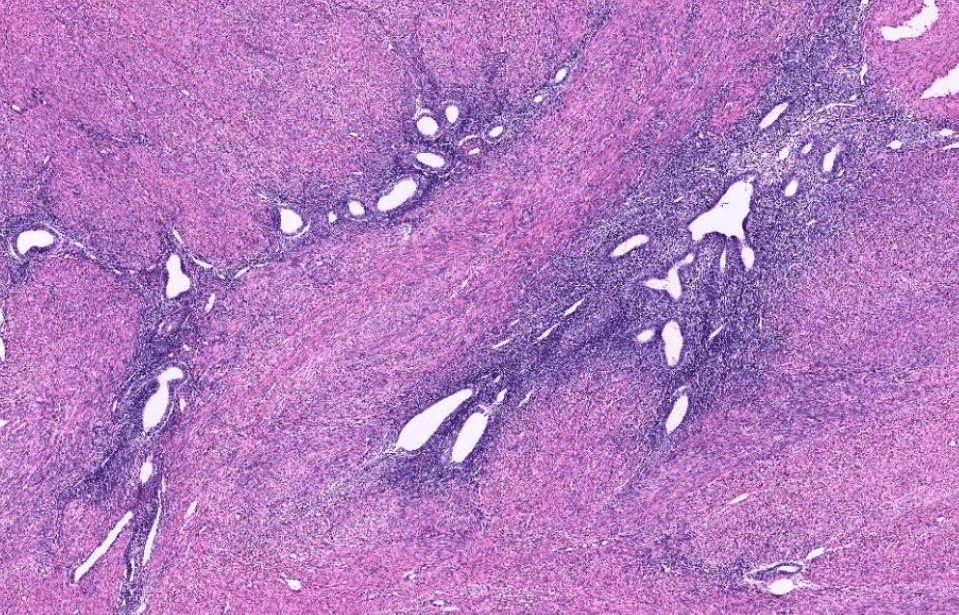
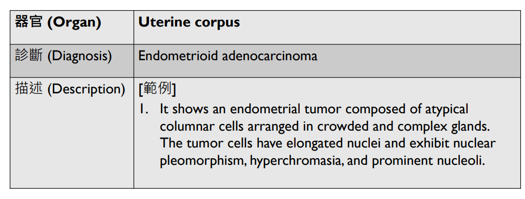
1. infiltrative/non-infiltrative endometrial tumor
2. atypical columnar cells arranged in crowded and complex glands, loss of intervening stroma
3. hyperchromatic elongated pleomophic nuclei, prominent nucleoli
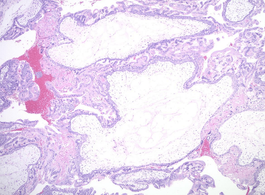
1. Avascular hydropic villi with central cisterns
2. Diffuse trophoblast hyperplasia with focal cytologic atypia
3. Endometrial decidual change and Arias-Stella reaction
唯一有villi的!
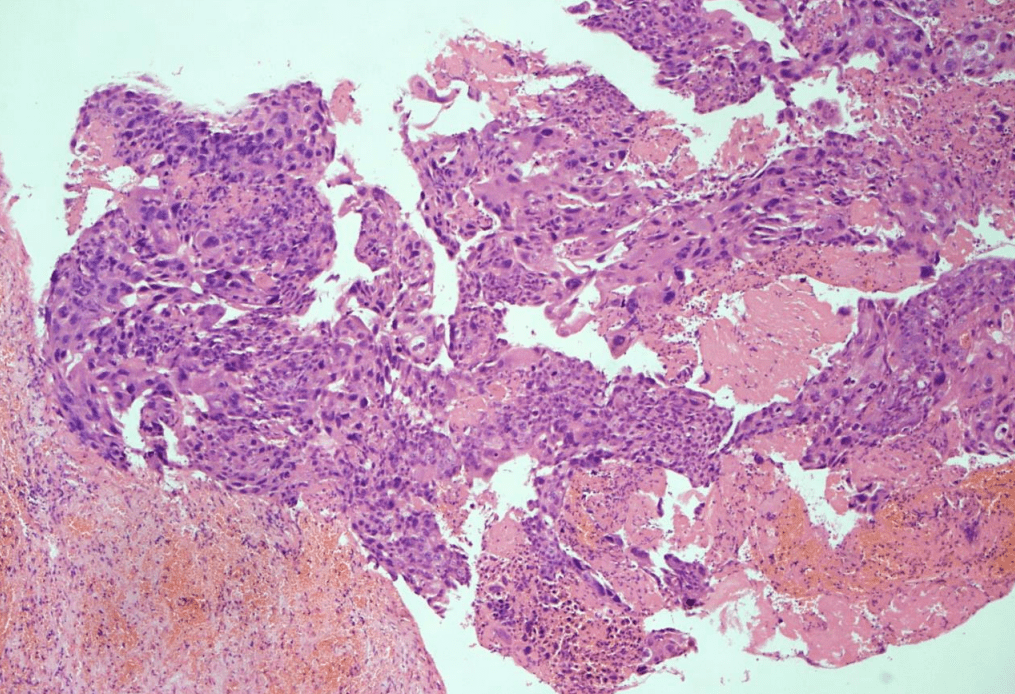
1. Infiltrative tumor invading the myometrium.
2. solid sheets of atypical multinucleated syncytiotrophoblasts, mononuclear cytotrophoblasts, and intermediate trophoblasts.
3. Marked cytologic atypia: hyperchromatic enlarged pleomorphic nuclei, increased mitosis.
4. may have necrosis/hemorrhage
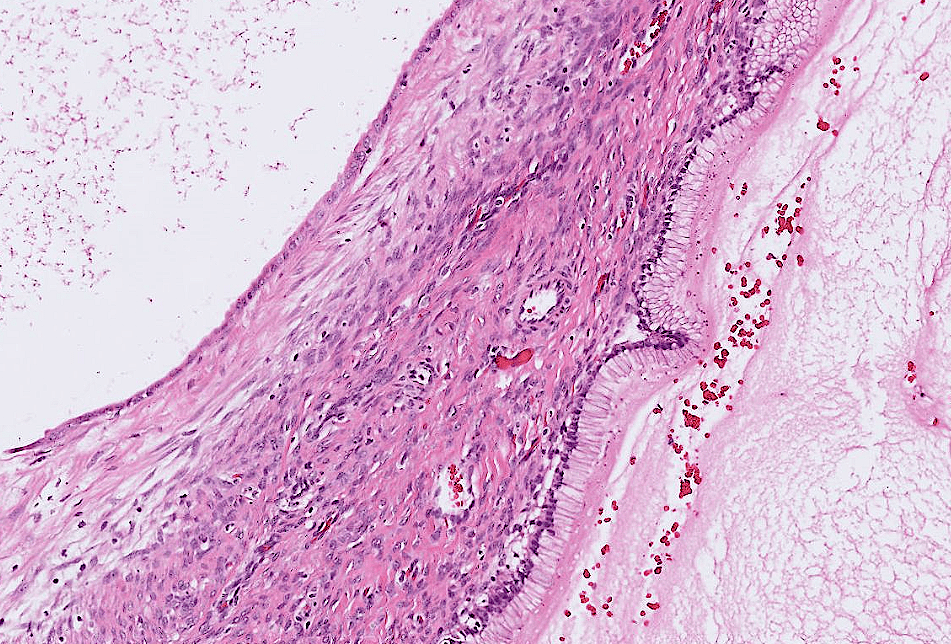
1. Multilocular cystic lesion lined by a single layer of tall columnar cells with abudant apical mucin
2. No definite cytologic atypia, stratification, papillary formation, or stromal invasion.
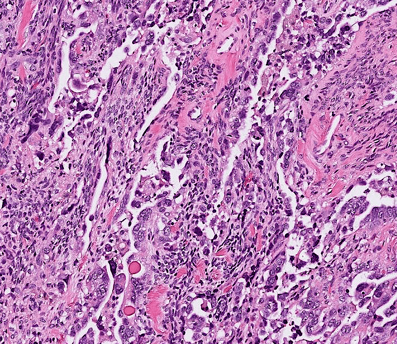
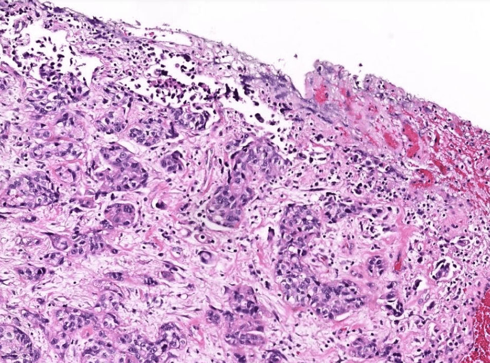
1. Infiltrative border
2. Papillary and solid growth of columnar to cuboidal tumor cells with eosinophilic cytoplasms.
3. Slit-like spaces between papillae.
4. Marked cytologic atypia: pleomorphic nuclei, prominent nucleloli, and frequent mitosis
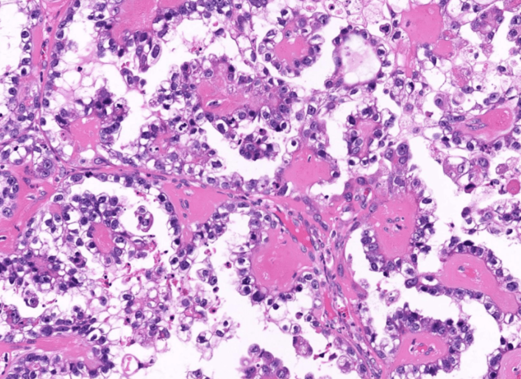
1. Tubulocystic and papillary structures lined by cuboidal, polygonal, or round cells with clear cytoplasm.
2. Papillae have hyalinized fibrovascular core.
3. Cells with hobnail-appearance.
4. Hyperchromatic pleomorphic nuclei, prominent nucleoli
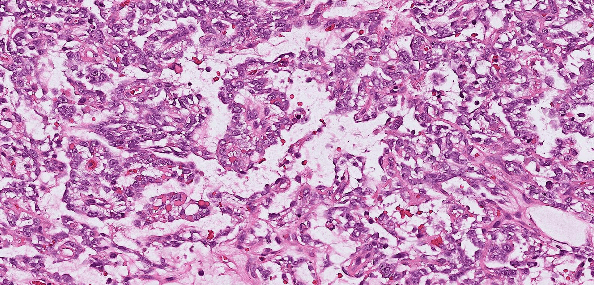
1. Well-defined tumor with mixed cystic / microcystic/ reticular pattern.
2. Cuboidal to flattened tumor cells with clear to pale eosinoplilic cytoplasm.
3. Schiller-Duval bodies and hyaline droplets.
4. Inconspicuous cytologic atypia.
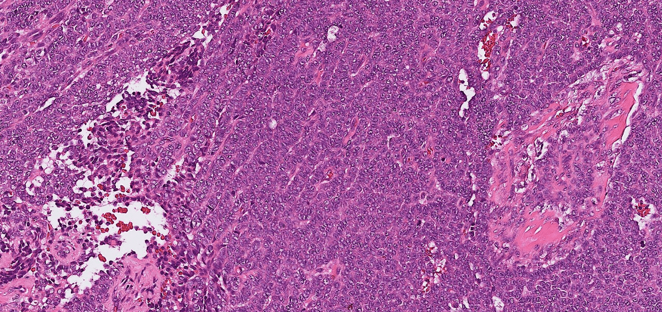
1. Well-defined tumor with mixed solid /trabecular /microfollicular patterns.
2. Call-exner bodies.
3. Uniform oval tumor cells with coffee bean-like grooved nuclei and scant eosinophilic cytoplasm.
4. Inconspicuous cytologic atypia.
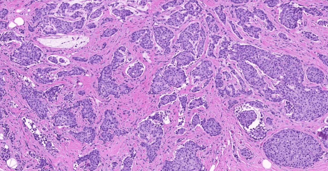
1. Infiltrative tubules.
2. Atypical cuboidal to columnar epithelial cells with hyperchromatic enlarged pleomorphic nuclei, prominent nucleoli, and increased mitosis.
3. cribriform (or other types of) DCIS
4. stromal desmoplasia
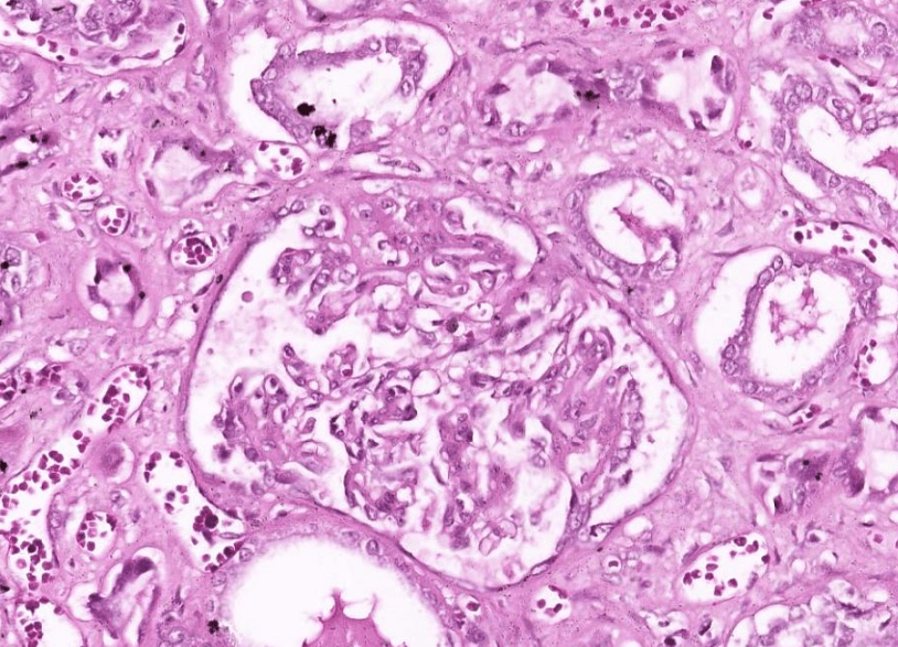
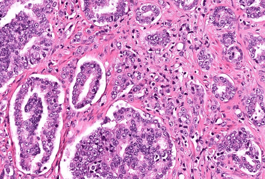
Organ: Kidney
Diagnosis: Focal Segmental Glomerulosclerosis
Discription:
1. Interstitial fibrosis/inflammation, tubular atrophy
2. some glomeruli show segmental sclerosis and adhesion to Bowman's capsule
3. arteriosclerosis
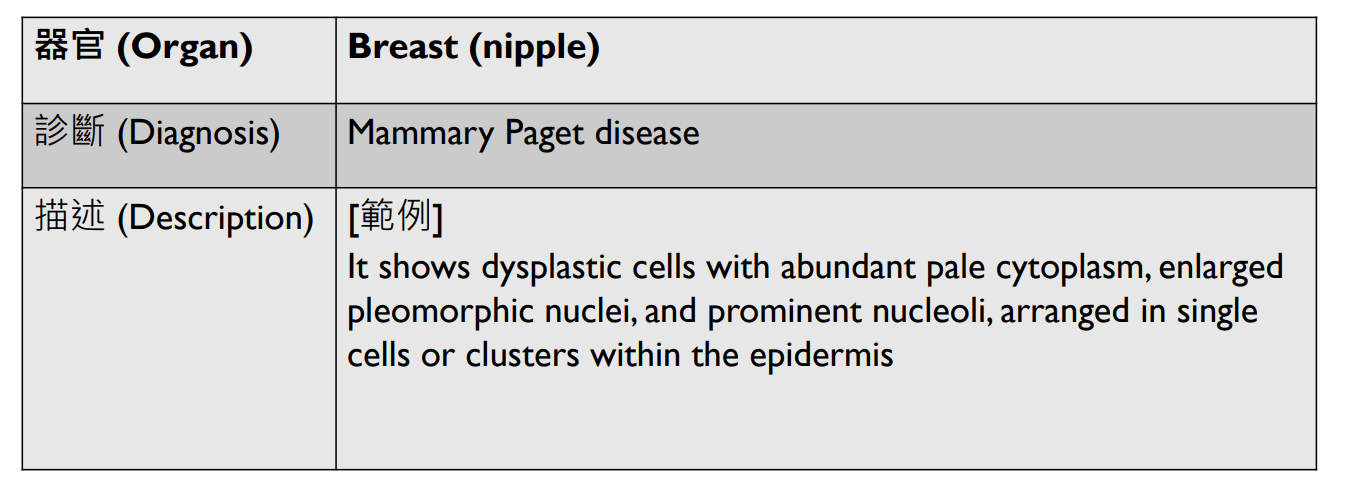
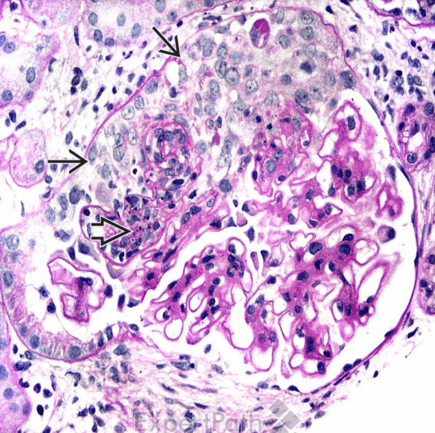
Organ: Kidney
Diagnosis: Diabetic Nephropathy
Discription:
1. interstitial fibrosis/inflammation, tubular atrophy
2. Diffuse mesangial sclerosis
3. Nodular glomerulosclerosis
4. hyaline arteriolosclerosis
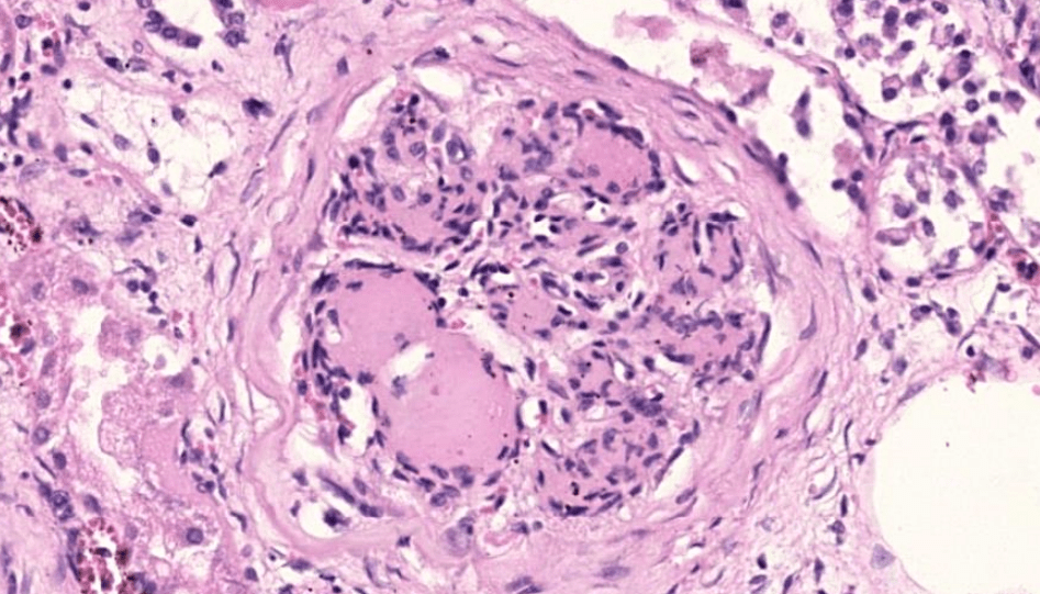
Organ: Kidney
Diagnosis: Class 4 lupus nephritis with crescents
Discription:
1. Hypercellular glomeruli with capillary wall thickening (wire loop)
2. crescent-shaped extracapillary proliferation
(3. interstitial fibrosis/inflammation, tubular atrophy)
題
By Zi-Hong Xiao


