Atul Jaidka PRO
Cardiologist | Unity Health - St. Joseph's Hospital
https://onlinelibrary-wiley-com.myaccess.library.utoronto.ca/doi/full/10.1111/echo.15945
https://onlinelibrary-wiley-com.myaccess.library.utoronto.ca/doi/full/10.1111/echo.15945
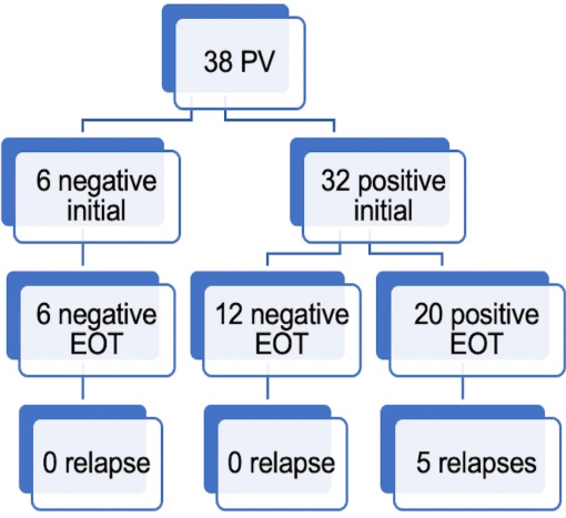
FDG Imaging:
Conclusion:
3 months later
FDG Imaging:
Conclusion:
By Atul Jaidka


