Cardiac Complications of Covid-19

Atul Jaidka

COVID-19

Review

  • COVID-19 is a coronavirus with host receptor ACE2
  • Person-person transmission through respiratory droplets
  • Incubation period mostly 4-5 days but as long as 14 days

Atri D, Siddiqi HK, Lang JP, Nauffal V, Morrow DA, Bohula EA. COVID-19 for the Cardiologist: Basic Virology, Epidemiology, Cardiac Manifestations, and Potential Therapeutic Strategies. JACC Basic Transl Sci. 2020; 5(5):518-536. [PDF]

Clinical Features

  • Pneumonia is most frequent manifestation
  • Symptoms:
    • fever, cough, dyspnea, myalgias, diarrhea, loss of smell and taste
    • no reliable clinical feature to reliably distinguish from other viral respiratory infections
  • ARDS is the major complication
  • Other complications:
    • Thomboembolic events, acute cardiac injury, kidney injury, inflammatory complications, and encephalopathy

Lab Features

UTD

Imaging Features

  • Chest radiographs:
    • normal, consolidation and ground-glass opacities
    • bilateral, peripheral, and lower lung zone distributions
  • Chest CT:
    • Ground-glass opacifications – 83 percent
    • Ground-glass opacifications with mixed consolidation – 58 percent
    • Adjacent pleural thickening – 52 percent
    • Interlobular septal thickening – 48 percent
    • Air bronchograms – 46 percent
  • Both Xray and CT may be normal on initial presentation and normalization may lag behind clinical improvement

Diagnosis

UTD

COVID-19

Cardiac Complications

Cardiac Complications

  • Myocardial Injury
    • Myocarditis
    • Stress cardiomyopathy
    • Myocardial infarction
  • Heart Failure
    • Right heart failure
    • Cardiogenic shock
    • Multisystem inflammatory syndrome in adults (MIS-A)
  • Cardiac arrhythmias

Evaluation

Recommended in all patients:

  • Troponin
    • may have prognostic value and useful as baseline
    • does not indicate ACS in absence of symptoms/ECG changes
  • ECG
    • baseline QRS-T morphology
    • QTc for potential future treatment
    • Reported abnormalities: T-wave depression/inversion, ST elevation and depression, and Q waves

Targeted Evaluation

Recommended in patients with new HF, ECG changes, or cardiac arrhythmias. Important considerations:

  • Likelihood will change management or guide prognosis
  • Factoring in nosocomial infection/spread
  • Managing limited medical staff resources

All patients:

  • Hx, Phx, ECG, Tn, BNP
  • Depending on clinical concern for ACS, stress cardiomyopathy, myocarditis, or heart failure further workup

Myocardial Injury

Myocardial Injury

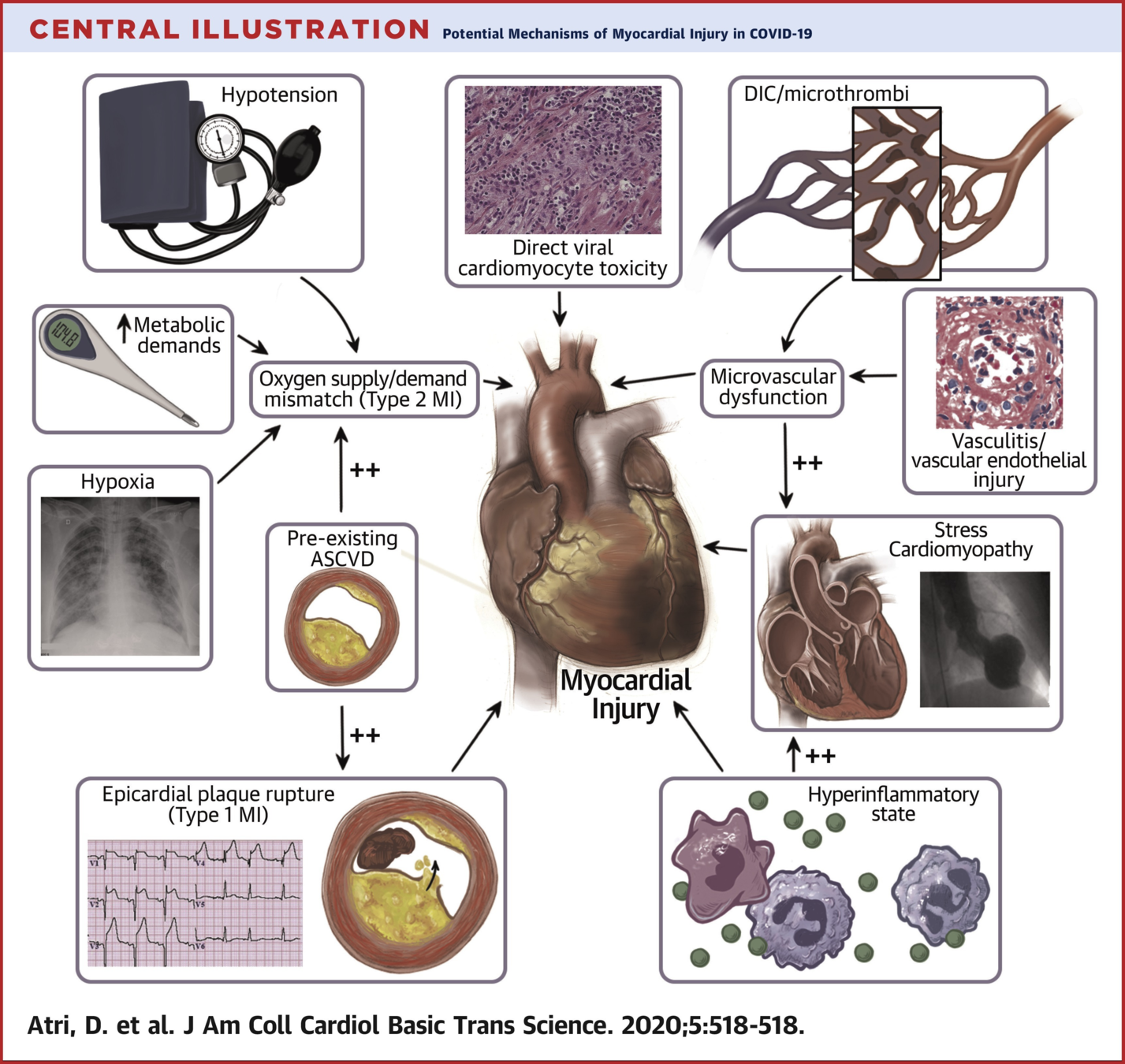
Cause unknown but proposed mechanisms:

  • Myocarditis
  • Stress cardiomyopathy
  • Ischemic injury from cardiac microvascular dysfunction
  • Small vessel cardiac vasculitis
  • Endotheliitis
  • Epicardial CAD (plaque rupture or demand ischemia)
  • Right heart strain (Lung disease or PE)
  • ARDS
  • SIRS, cytokine storm


Unknown if ACE2 signaling pathway has a role in COVID-19 cardiac injury

Myocarditis

Myocarditis

  • Many case reports with diagnosis of clinically suspected myocarditis but few biopsy confirmed cases.
  • Very few patients have been definitely diagnosed with COVID-19 myocarditis (COVID-19 infiltrate detected in the myocardium)
  • If myocarditis suspected, and diagnosis with change management/prognosis, recommend:
    • Echocardiogram (LV/RV dysfunction or dilation)
    • MRI (non-specific findings, published studies not correlated to biopsy)
    • Endomyocardial Biopsy (helpful to rule out other treatable causes of myocarditis such as giant cell)

Definitive Diagnosis of Myocarditis?

  • Dallas Criteria:
    • Active myocarditis: inflammatory infiltrate of the myocardium not typical of ischemic damage

Myocarditis

  • No targeted treatment
  • Treat LV Dysfunction
  • ACE and ARBs are safe

Stress Cardiomyopthy

Criteria:

  • Transient left ventricular (LV) systolic dysfunction (typically not in a single coronary distribution; patterns include apical, midventricular, and basal).
  • Absence of angiographic evidence of obstructive coronary disease or acute plaque rupture (or if coronary artery disease is present, the wall motion abnormalities are not in the territory of the affected coronary artery).
  • New ECG abnormalities (ST-segment elevation and/or T-wave inversion) or modest elevation in cardiac troponin.
  • Absence of pheochromocytoma or myocarditis.

COVID-19 Patients

  • Case reports in patients with COVID-19
  • Review of 12 cases:
    • Mean age 70.8
    • Mostly female
    • Elevated troponin
    • Complications include HF, cardiogenic shock, cardiac tamponade, hypertensive crisis
  • Typically recovers in 1-4 weeks
  • Treat as usual takotsubo

Myocardial Infarction

Myocardial Infarction

  • CAD common in patients with COVID-19 (4.2-25%)
  • Unclear if ACS in COVID-19 patients is due to direct or indirect cause
  • ACS patients the suspicion, treatment, and management as usual
    • Can consider liberalizing usage of fibrinolytic therapy
  • Education to public to seek care for ACS
  • Stable CAD patients also treated the same except consider delaying revasc if indication is relief of symptoms

Heart Failure

Heart Failure

  • Causes:
    • Precipitated by acute illness in patients with known or undiagnosed heart disease
    • Acute hemodynamic stress (ie. acutely elevated right sided pressures)
    • Acute myocardial injury (differential as previous discussed)
  • Associated with worse outcomes
  • Incidence unknown but appears increased in COVID-19 patients

Right Heart Failure

  • Acute cor pulmonale: right heart failure due to acutely rising pulmonary pressures
  • Causes:
    • PE, ARDS
  • Treat as usual
    • Underlying cause (ie. anticoag)
    • Volume management
    • Inotropes
    • Pulmonary vasodilation

Cardiogenic Shock

  • Case reports
  • Unclear cause, myocarditis suspected but not definitely diagnosed
  • Usual management with intropes and vasopressors
  • Reserve mechanical support for INTERMACS 1-3
  • Waitlist on transplant list until 14 days post diagnosis and 2 negative swabs

Classification of Cardiogenic Shock

Cardiac Arrhythmias

Cardiac Arrhthmias

  • Vast majority of patients do not have arrhythmia related signs/symptoms
  • Arrhythmias may occur in patients with myocardial injury, myocardial ischemia, hypoxia, shock, electrolyte disturbances, or those receiving QT prolonging drugs
  • 12 lead ECG in all patients
  • If QTC >500 or 60ms above baseline, correct lytes and tele
  • Torsade des pointes (TdP) treated as per usual
    • cardioversion/defib
    • correct electrolytes
    • remove drugs
  • ACLS as usual but with precautions (ie. protected code blue)

Questions?

Cardiac Complications of Covid-19

By Atul Jaidka

Cardiac Complications of Covid-19

  • 28