Background

ID: 31M admitted for SOB


PMH: Remote IVDU, Endocarditis 2011 of AV/MV complicated by severe AR resulting in Prosaic Bioprosthetic Valve inserted, residual significant MR


MEDS: Carvedilol, Candarsartan, Lasix


Social: No more IVDU


HPI: 2 week history of SOB, cough, subjective fevers/chills, NYHA Class 3-4.

Background

Exam: VS stable, no oxygen requirements, afebrile, mild clinical hypovolemia


Previous Echo:

  • TTE/TEE 6 months previous: Flail Ant MV with perforation at base of leaflet, severe MR, chronically elevated prosthetic aortic valve gradients, no masses or vegetations


Plan:

  • Admitted to reassess valvular disease given significant shortness of breath on exertion

Previous TTE

Current TTE

Previous

TTE

  • Normal left ventricular size and function.
  • Normal right ventricular systolic function.
  • Anterior paravalvular abscess extending into the ventricular septum. Abnormal colour flow into the right ventricle, suspicious for septal perforation through the abscess cavity into the right ventricle.
  • Aortic prosthesis gradients severely elevated
  • A non-mobile echodensity is noted on atrial aspect of the anterior mitral leaflet, consistent with vegetation.​

Investigations

Blood cultures: Negative (remained afebrile)


CT Abdo: Splenic infarct


CT Head: Frontal subarachnoid hemorrhage with associated mycotic aneurysm

CT Heart

CT Heart

Presumed paravalvular abscess adjacent to the bioprosthetic aortic root including communication with the subvalvular LVOT. Multiple areas of low attenuation within the cavity, the right ventricle and the mitral valve likely represent vegetations versus thrombus.

Contrast

Contrast TTE

Moderate size mobile filling defect at the right ventricular apex, not seen on non-contrast images,
suspicious for chordal based vegetation or thrombus.
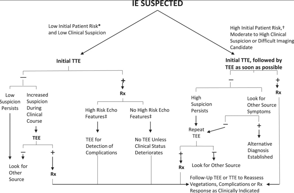
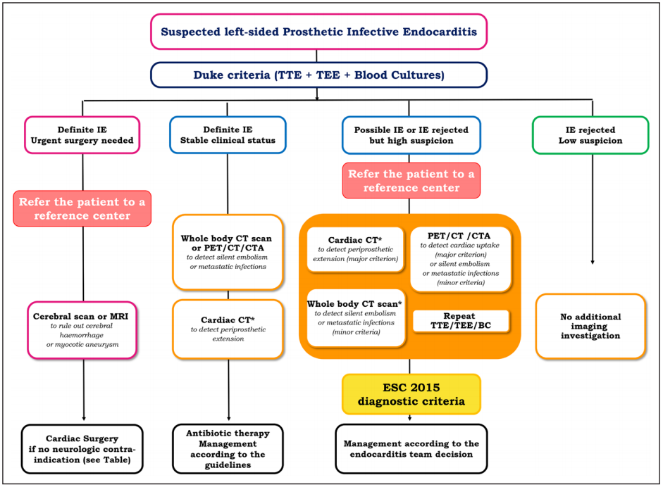
TEE

TEE

  • Large echolucent space anterior to the aortic prosthesis extending into the basal ventricular septum. Colour flow noted into this space at the level of the bioprosthesis with diastolic flow from this presumed abscess cavity into the left ventricular outflow tract, consistent with paravalvular aortic abscess with rupture into the left ventricular outflow tract.


  • Large mass on the atrial aspect of the anterior mitral valve, extending into the left atrium, consistent with vegetation.  

Discussion

Aortic Perivalvular Abscess

  • Reported in 30-40% of patients with IE
    • Most commonly in aortic valve infection, especially when located between right and non-coronary cusp
    • Mitral valve and annulus less common
  • Clues:
    • conduction abnormalities
    • persistent bacteremia
    • persistent fevers
  • Risk Factors: possibly vegetation size, bicuspid valve, and intravenous drug use
    • Our patient did not have any of these clues or active risk factors

Aortic Perivalvular Abscess

  • Can invade into neighbouring tissue
    • Commonly into conduction tissue causing heart block
    • Rarely can cause coronary compression and acute coronary syndrome
  • In this case very uncommon to invade through septum and cause right ventricular chordal based infection
  • Clinically important as associated with increased risk of systemic embolization (twice as high in some studies) and worsening mortality

Aortic Perivalvular Abscess

  • TEE more sensitive for detection for myocardial abscess than TTE
  • Specificity high in TTE but should still move to TEE to assess for extent of involvement

AHA

Mutlimodality Imaging

  • Other imaging for endocarditis includes Cardiac CT, MR, WBC-SPECT, and PET
  • CT: helpful for paravalvular infection and but can miss small vegetations
    • In this case was important for preoperative planning but also allowed for picking up on infection that was otherwise missed on TTE
  • Some propose an Imaging Team within the Endocarditis Team 

Proposed Imaging Algorithm

Erba PA, Pizzi MN, Roque A, et al. Multimodality Imaging in Infective Endocarditis: An Imaging Team Within the Endocarditis Team. Circulation. 2019; 140(21):1753-1765.

Case Discussion

  • Endocarditis can be aggressive thus need to use all tools available to completely visualize lesions before OR (TTE/TEE/Contrast/CT)
    • Repeat imaging helpful to build off on previous information and further visualize lesions/connections
  • Despite being culture negative and afebrile, endocarditis still possible, especially if subacute onset
    • High index of suspicion in patients with prosthetic valves or pre-existing disease
  • Visualizing heart in 3D helpful to conceptualize lesions that span between views

Case Resolution

Management Challenge

  • Balancing risk of subarachnoid hemorrhage and urgent need for cardiac surgery where anticoagulation would be used
  • Serial CT Head completed and once cleared by Neuro Sx, proceeded with OR


OR:

  • Aortic valve explantation and extensive debridement of aortic root and fibrous trigones.
  • Aortic root replacement (26 mm LifeNet aortic homograft).
  • Palliative, complex mitral repair (A2 triangular resection, debridement of posterior medial commissure, A1 perforation closure and 34 mm Cosgrove band annuloplasty).
  • Repair of atrioventricular groove, reconstruction of fibrous trigones.
  • Patent foramen ovale closure.


Follow-up:

  • Cultures remained negative. Finished 6 weeks of antibiotics
  • 1 year post OR: feels well,  NYHA 1

Thank You

Questions?

Western Echo Symposium - Interesting Case

By Atul Jaidka

Western Echo Symposium - Interesting Case

Echo Rounds - Jan 22

  • 31