

Atul Jaidka PRO
Cardiologist | Unity Health - St. Joseph's Hospital
Atul Jaidka
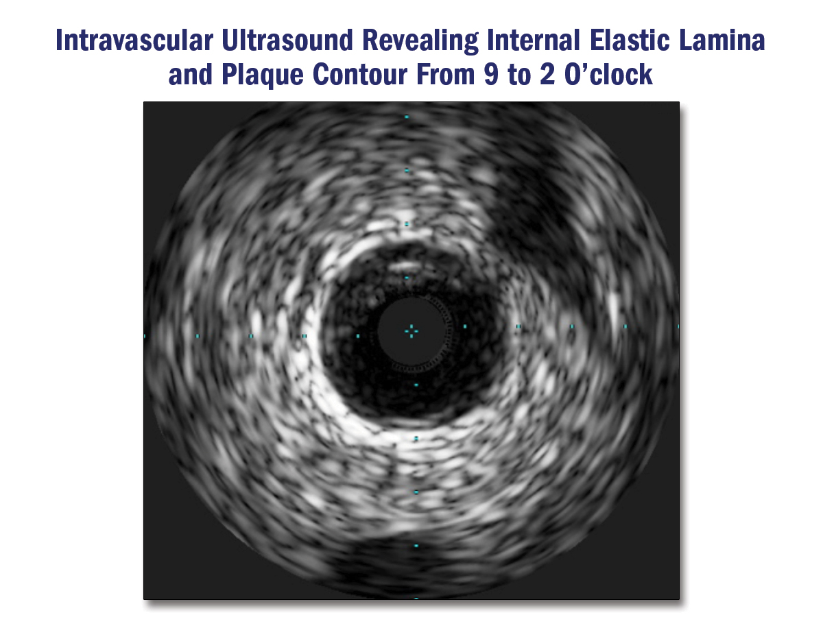
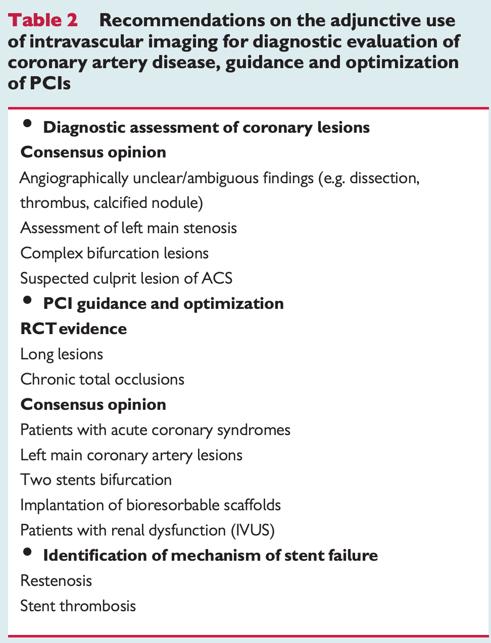
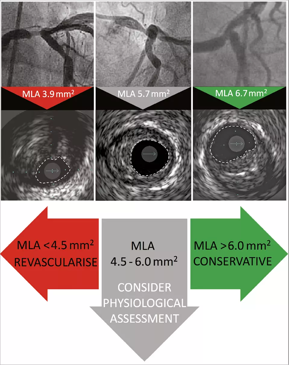
In patients with intermediate stenosis of the left main artery, intravascular ultrasound (IVUS) is reasonable to help define lesion severity (Class IIa).
In patients with intermediate stenosis of the left main artery, intravascular ultrasound (IVUS) is reasonable to help define lesion severity (Class IIa).
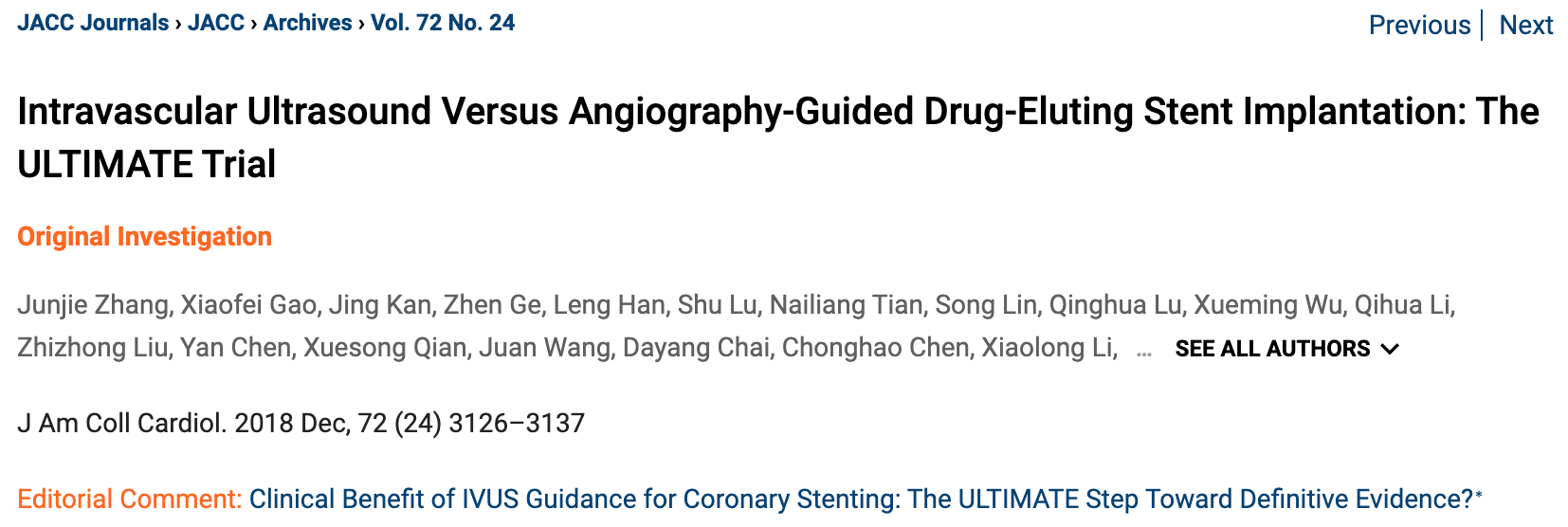
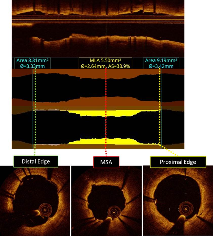
In patients undergoing coronary stent implantation, IVUS can be useful for procedural guidance, particularly in cases of left main or complex coronary artery stenting, to reduce ischemic events (Class IIa).
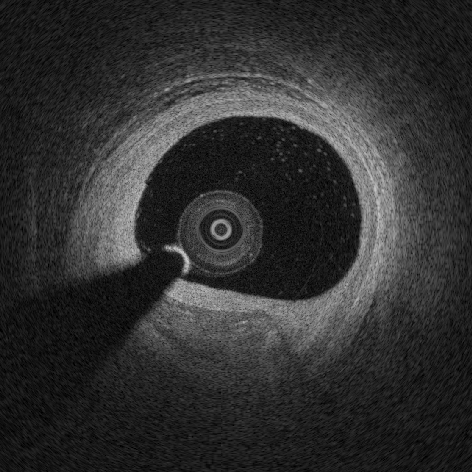
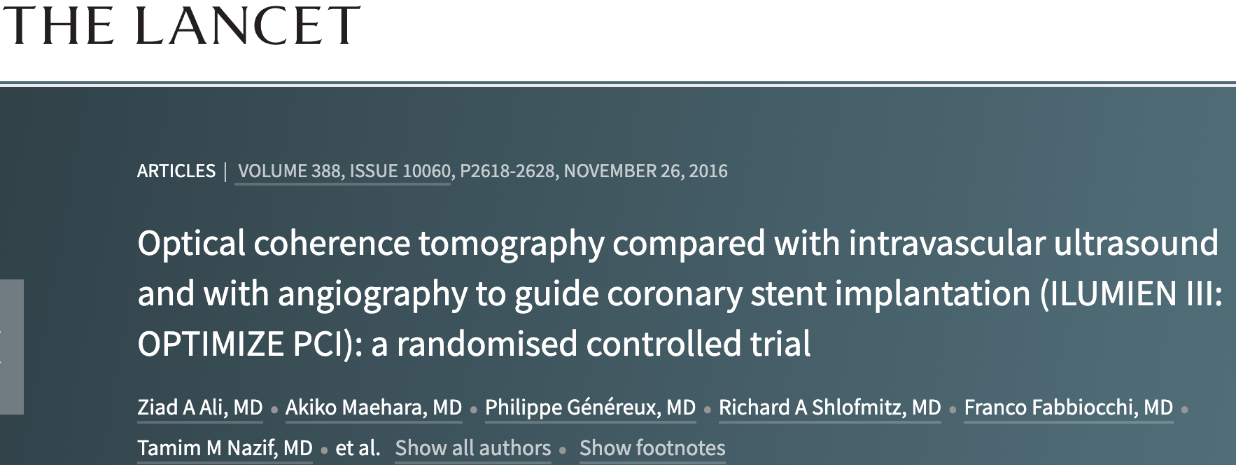
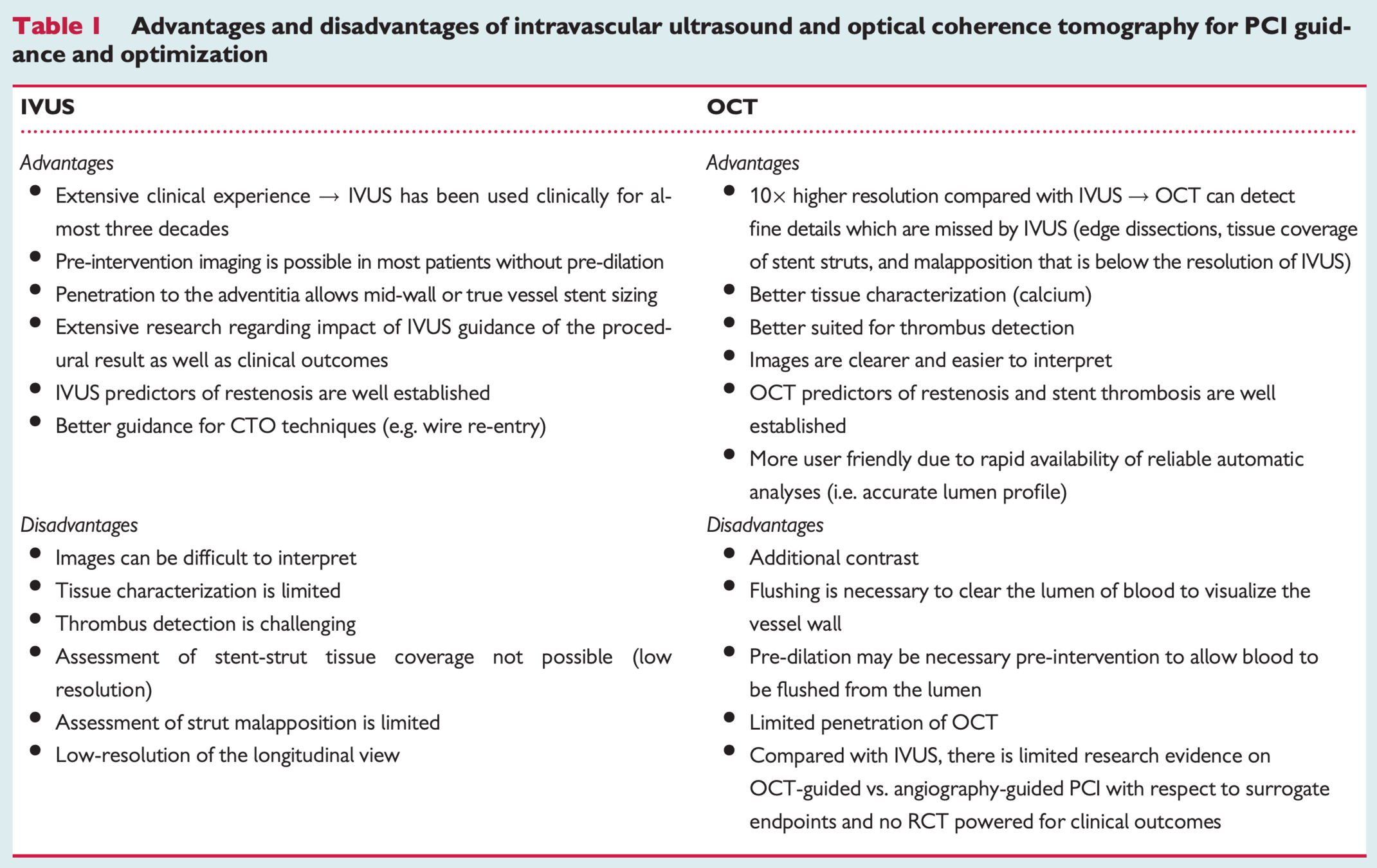
In patients undergoing coronary stent implantation, OCT is a reasonable alternative to IVUS for procedural guidance, except in ostial left main disease (Class IIa).
https://academic.oup.com/eurheartj/article/39/35/3281/5001185
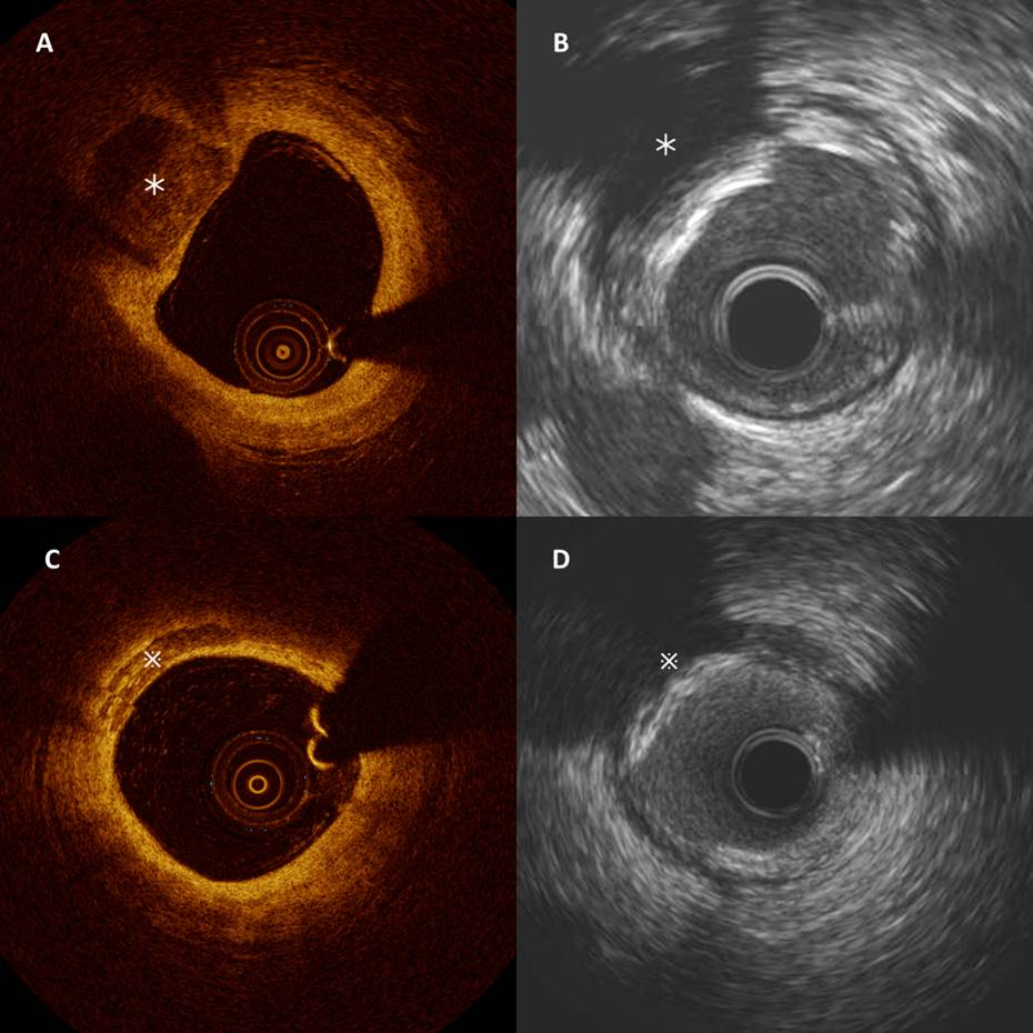
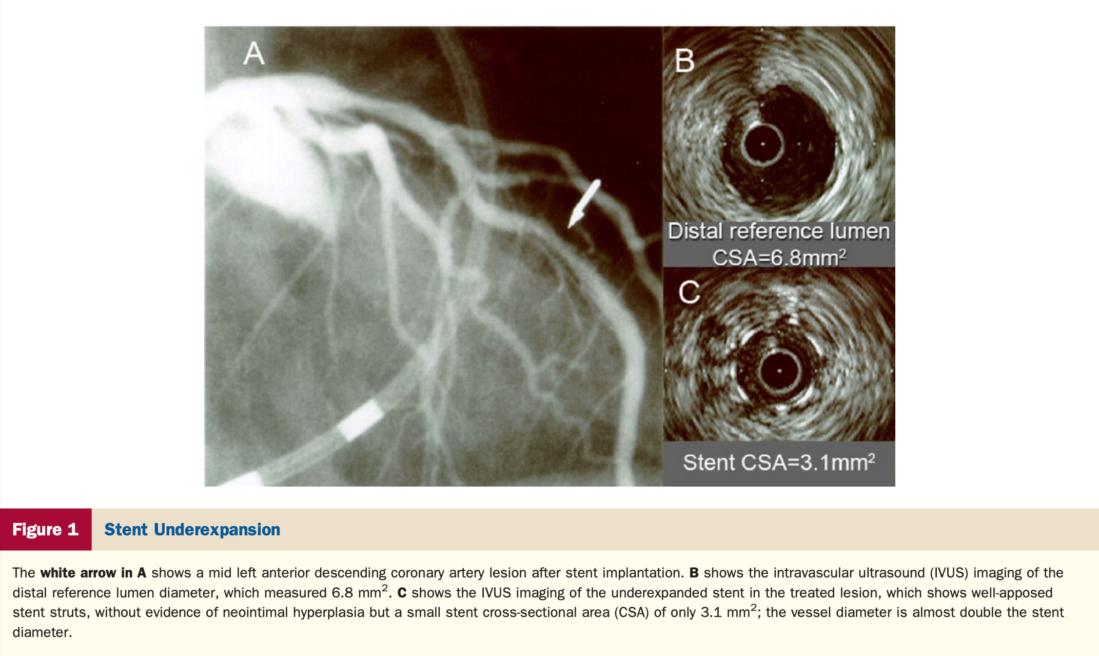
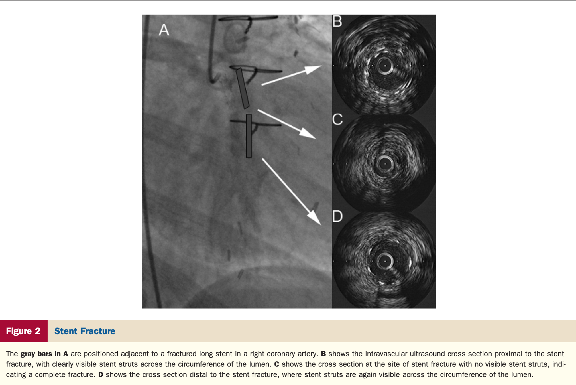
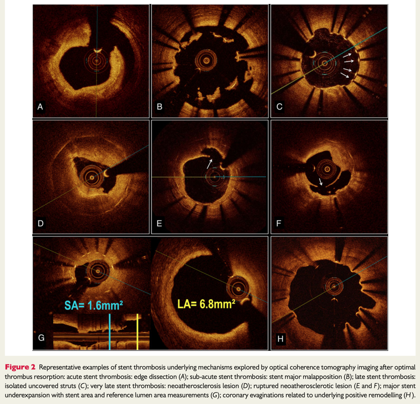
In patients with stent failure, IVUS or OCT is reasonable to determine the mechanism of stent failure (Class IIa).
In patients with stent failure, IVUS or OCT is reasonable to determine the mechanism of stent failure (Class IIa).
https://academic.oup.com/eurheartj/article/40/31/2566/5491794
https://www.acc.org/latest-in-cardiology/articles/2016/06/13/10/01/intravascular-oct-in-pci
https://www.acc.org/latest-in-cardiology/articles/2016/06/13/10/01/intravascular-oct-in-pci
https://www.acc.org/latest-in-cardiology/articles/2016/06/13/10/01/intravascular-oct-in-pci
https://www.jacc.org/doi/abs/10.1016/j.jacc.2010.07.028
https://www.jacc.org/doi/abs/10.1016/j.jacc.2010.07.028
https://academic.oup.com/eurheartj/article/37/15/1208/1748814
By Atul Jaidka


