Pericardial Diseases

Atul Jaidka | Echo Rounds

Objectives

  • Anatomy
  • Tumours
  • Pericardial Cyst
  • Congenital
  • Pericardial Fat
  • Pericardial Effusion
  • Tamponade
  • Pericarditis
  • Constriction

Case!

Code Stemi

  • Cath: Non-obstructive CAD
  • Admitted to CCU
  • High O2 Requirements
  • Please do echo for HF

Conclusion

  • History of laryngeal SCC
  • CT also shows pulmonary nodules
  • Biopsy pending to confirm diagnosis
  • Likely Metastatic SCC

Anatomy

Anatomy

  • Double layers structure with visceral and parietal
  • Visceral (serosal) is attached to epicardium and parietal (fibrous) surrounds the heart
  • Normally 20-50mL of fluids present

Echo Protocol

Echo Protocol

  • 2D echocardiography
    • PEff (size, location, free flowing vs organizing, suitability for pericardiocentesis vs window)
    • Pericardial thickness (particularly TEE)
    • Collapse of right-sided chambers (duration of diastole and relation with respiration)
    • Early diastolic septal bounce, respiratory shift of the ventricular septum
    • IVC plethora
    • Pleural effusion/ascites
    • RA tethering (best seen by TEE)
    • Stasis of agitated saline contrast in right atrium (sluggish flow)

Echo Protocol

  • Doppler with Respirometry
    • Restrictive mitral inflow pattern
    • Reciprocal respiratory changes of mitral (and tricuspid) inflow
    • Reciprocal respiratory changes of diastolic forward flow velocity and end diastolic flow reversal in hepatic veins
    • Tissue Doppler velocities of mitral annulus, color Doppler M-mode of mitral inflow
    • 2D strain of longitudinal and circumferential deformation
  • M-Mode
    • Flattening of the posterior wall during diastole
    • Respiratory variation of ventricular size

Tumours

Pericardial Masses

https://www.acc.org/latest-in-cardiology/articles/2016/07/12/13/06/cmr-and-pericardial-masses

  • Masses can be from primary tumour, metastases, or mass encroaching on pericardium
  • Almost always effusion
  • Metastatic disease most commonly to pericardium

Case - Pericardial Lipoma

https://www.ncbi.nlm.nih.gov/pmc/articles/PMC5128889/

Pericardial Cysts

Pericardial Cyst

  • Usually along right heart border
    • can be anywhere
  • Generally asymptomatic unless compressing structure (ie coronary)
  • Important to distinguish from mass
  • Appear as echo free space, localized and spherical (vs pericardial effusion)
  • Further imaging needed

UTD

Case

https://academic.oup.com/ehjcimaging/article/12/11/E43/2396998

Case

https://academic.oup.com/ehjcimaging/article/12/11/E43/2396998

Case

https://academic.oup.com/ehjcimaging/article/12/11/E43/2396998

Congenital Absence of Pericardium

Congenital Absence of Pericardium (CPA)

  • Pericardium stabilizes the heart and limits dilation
  • Absence of pericardium is rare
  • Can be complete bilateral (9%), complete left (70%), complete right (17%), or partial (3-4%)
    • Partial can be life threatening due to possible herniation  (strangulate cardiac structure or compress coronaries)
  • 50% of cases have associated findings
    • ASD, PDA, ToF, etc
  • Complete usually asymptomatic
  • Partial can have trepopnea (dyspnea on one side or chest pain (compression of coronaries)

ECG

  • Normal in complete
  • Left CPA can have RAD, RBBB, poor transition

CXR

  • Left CPA heart can shift posterior and left causing straightening and elongation of heart (levorotation and levoposition)
  • "Snoopy" Sign

ECHO

  • Complete CPA better imaged supine give heart shift posterior and leftward
  • Exaggerated movement of heart in chest
  • Outward bulging of LV inferior wall seen best on 2 Chamber (tear drop shape)
  • Need further imaging to confirm
    • CMR gold standard

Case 1

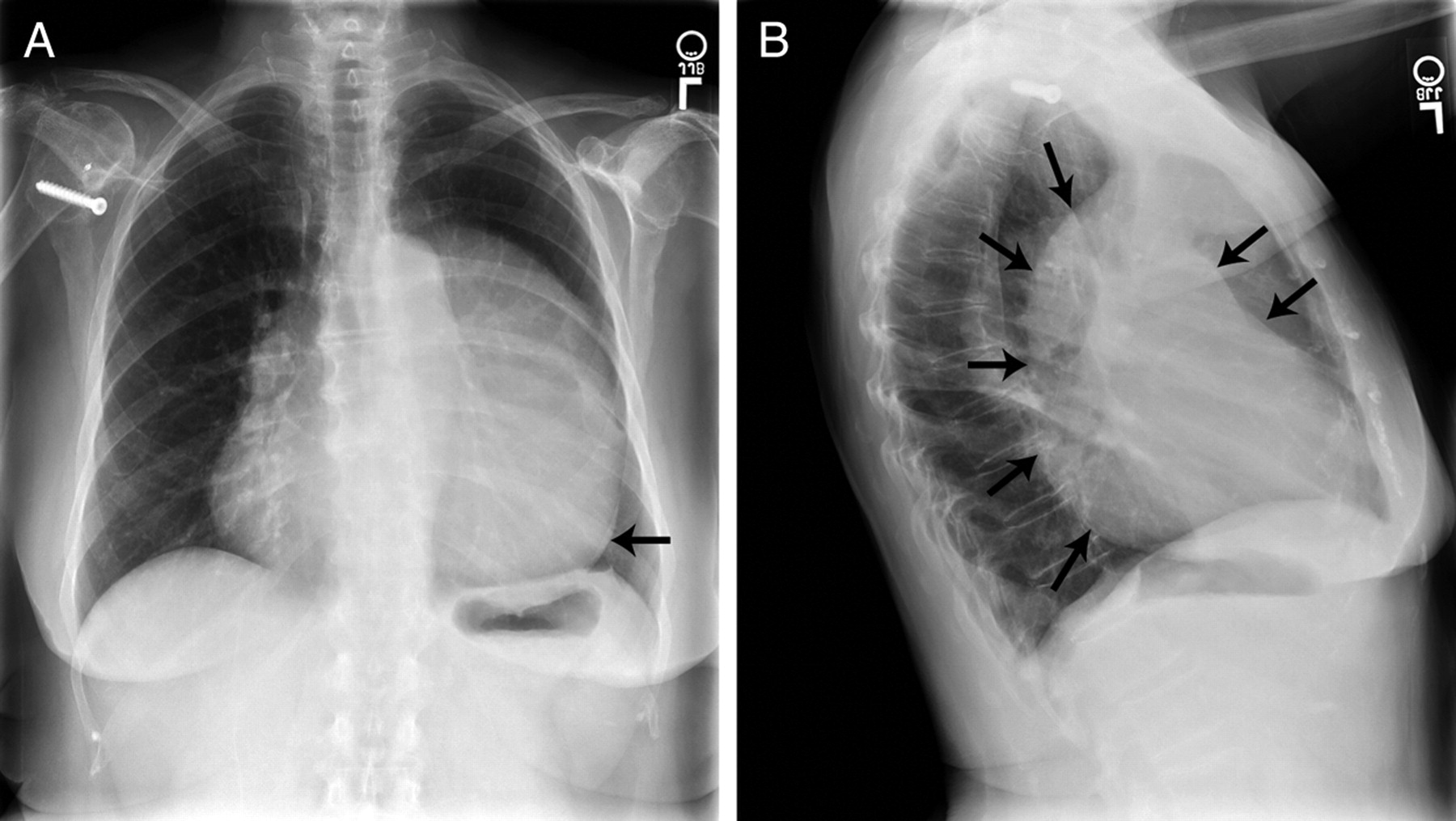
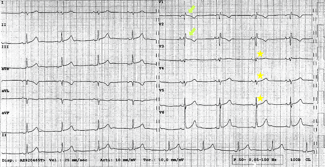
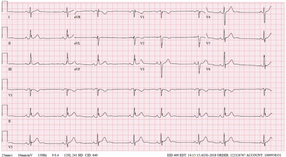
ECG: incomplete RBBB, poor R wave progression, right axis deviation

29M presented with atypical chest pain
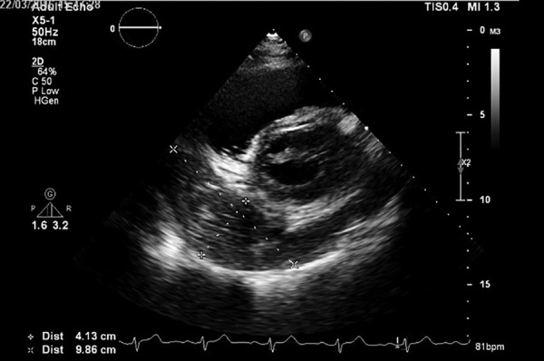
Case 1

A4C Medial

A4C Lateral

Note posteriorly directed apex and exaggerated mobility. "Teardrop" ventricles. Usual apical location shows RV predominance requiring supine positioning

https://www.cvcasejournal.com/article/S2468-6441(19)30165-3/fulltext

Case 2

Right ventricle appears enlarged, teardrop-shaped heart, and hypermobile heart

https://www.cvcasejournal.com/article/S2468-6441(19)30165-3/fulltext

"Pericardial Fat"

https://www.ncbi.nlm.nih.gov/pmc/articles/PMC3998169/

  • Different embryological origins
  • Epicardial fat has similar origin to omental and mesenteric fat
    • Correlates with BMI and metabolic syndrome
    • Better surrogate for cardiac disease
  • Epicardial fat (not paracardial fat) shared common blood supply with myocardium
    • Thought to have paracrine properties and cause for myocardial inflammation
    • Autopsy studies show epicardial fat extending into myocardium

Epicardial and Paracardial Fat

https://www.jacc.org/doi/pdf/10.1016/j.jacc.2011.05.052

Epicardial and Paracardial Fat

UTD

  • dashed arrow: "paracardial fat"
  • arrowhead: epicardial fat
  • arrow: visceral pericardium
  • thick arrow: parietal pericardium

Epicardial and Paracardial Fat

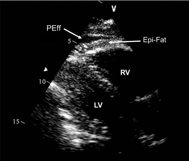
https://www.journal-of-cardiology.com/article/S0914-5087(14)00203-2/pdf#:~:text=On%20the%20other%20hand%2C%20the,(pericardi%2Dal%20fat).

Epicardial Fat vs Pericardial Effusion

  • Epicardial fat is often brighter than myocardium
  • Moves in concert with heart
  • Pericardial effusion is usually echolucent and motionless

ASE

Pericardial Effusion

Indications for Echo

  • Chest pain consistent with pericarditis or dissection
  • Enlarged cardiac silhouette on chest x-ray
  • Systemic disease associated with effusion
  • Post-MI
  • Hemodynamic instability after cardiac procedure

Sizing

  • Measure between visceral and parietal pericardium at end diastole (multiple spots to increase accuracy to assess volume)
  • Trivial (<10mm) <50mL
    • seen only in systole
  • Small (<10mm) 50-100mL
  • Moderate (10-20mm) 100-500mL
  • Large (>20mm) >500mL

Examples

ASE

Pleural Effusion

  • Assess on PLAX view
  • Fluid between descending aorta and heart is pericardial
  • Fluids below descending aorta is pleural

ASE

Stranding

UTD

  • Pericardial stranding suggests fibrous material in the pericardial effusion
  • Commonly seen post cardiac surgery, pericardial hemorrhage and in inflammatory process

Hematoma/Loculated Effusion

UTD

  • Large pericardial clot causing obliteration of right atrium

Tamponade

Tamponade - Chamber Collapse

  • RA and RV diastolic chamber collapse most commonly seen
  • RA collapse sensitive not specific
    • If exceeds 1/3 cardiac cycle very sensitive and specific
  • No chamber collapse has 90% negative predictive value

Tamponade - Resp Variation

  • Resp Variation
    • Formula: (Exp-Insp)/Exp
      • Measure first beat after insp and exp
    • Mitral >30%
    • Tricuspid >60%
  • Should not be used in isolated
  • Differentiate from COPD:
    • Tamponade first insp beat largest vs COPD second beat

Acute Pericarditis

Acute Pericarditis

  • All patients should undergo echocardiography
  • Assess for:
    • Pericardial effusion
    • Exclude other etiologies of chest pain

Constriction

  1. Ventricular septal motion abnormality (from ventricular interdependence)
  2. Medial mitral annulus e' velocity ≥ 9 cm/sec
  3. Hepatic vein expiratory diastolic reversal ratio ≥ 0.79 (Figure)
  4. Restrictive mitral inflow velocity (E/A ratio > 0.8)
  5. Plethoric inferior vena cava

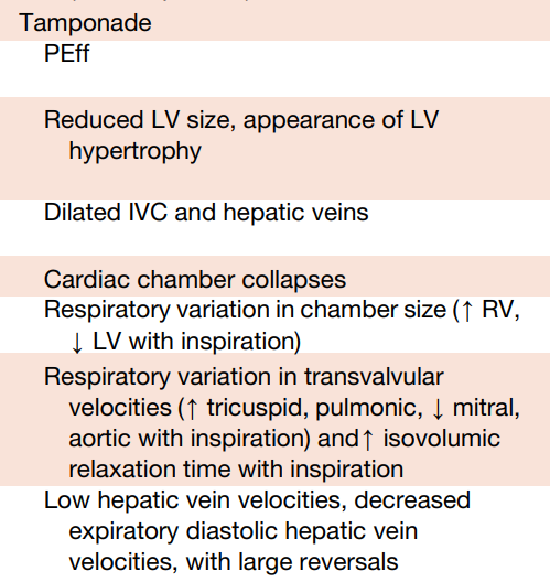
1 + either 2 or 3 =  87% sensitive and 91% specific

Diagnosis

Pericardial Diseases

By Atul Jaidka

Pericardial Diseases

Echo Rounds

  • 35